Photo Atlas of Ocular DiseaseReview of Optometry has created the following new resource—a photo atlas of ocular diseases across the entire spectrum of eye care. In addition to over 200 high-quality photos, you'll find clinical pearls for each condition and links to relevant articles that provide detailed guidance. An index of the entire atlas can be found here or you can check out the conditions featured in their respective categories: |
As primary eyecare providers, optometrists are responsible for the evaluation, management and, in many cases, the in-office treatment of a wide variety of eyelid lesions. Evaluation of the lids and adnexa is an important part of any eye exam and for us that begins with our initial face-to-face encounter with the patient. General characteristics such as symmetry (both inter-eye and intra-eye), skin color, loss of lashes, injection, bleeding and crusting are often evident by gross examination at a normal social distance.
A thorough slit lamp exam can then further inspect any areas of suspected pathology. It is important to look at the lids both closed and open to fully assess for any lesions and observe the apposition of the lids to the globe. In general, it is best to begin your slit lamp exam at a low magnification (6.3x or 10x) with a broad beam providing diffuse illumination. The anatomy of the lids, with their wide array of tissue and gland types, as well as the recognition that eyelid skin is typically uncovered and exposed to environmental factors such as UV light, explains the variety of lumps and bumps encountered in optometric practice.
Documentation of these lesions, including measurement of both vertical and horizontal meridians, will allow the clinician to monitor for growth or potential change to malignancy at future exams. Lesions chosen for in-office removal should be sent for pathologic evaluation, as research has shown that a small percentage of these that present with only benign characteristics are indeed cancerous. Those with malignant characteristics should be sent to a trusted oculoplastics ophthalmologist familiar with the appropriate comanagement of such conditions. When in doubt, it is wise to thoroughly document the lesion (photographic documentation is quite useful), educate the patient on malevolent signs of conversion to malignancy and ensure appropriate follow-up for clinical re-evaluation.
This section of photos should help distinguish eyelid/adnexal conditions that may have overlapping characteristics at presentation, as well as showcase more abnormal or rare presentations of frequent, common conditions. We hope to provide you a quick, convenient reference for all your eyelid evaluation needs!
Feel free to click the condition that interests you to jump to it, or scroll to look through all the conditions.
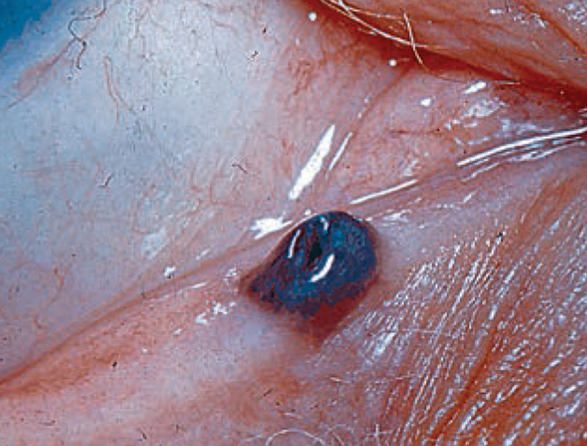
Junctional nevus
 |
| Photo: Rodney Bendure, OD, Jackie Burress, OD. Click image to enlarge. |
In junctional nevi, melanocytes proliferate in the epidermis at the epidermal-dermal border, eventually migrating to the dermis.
Suggested reading:
Don't Be Stumped by These Lumps and Bumps
Recognize Benign vs. Malignant Eyelid Tumors and Lesions
Xanthelasma
 |
| Photo: Rodney Bendure, OD, Jackie Burress, OD. Click image to enlarge. |
These lesions usually present on the eyelids as yellow plaques filled with lipid-laden macrophages. They arise after the age of 50 and should prompt suspicion of a lipoprotein disorder when present in patients younger than this. These may be surgically removed, but do tend to recur 50% of the time.
Suggested reading:
Don't Be Stumped by These Lumps and Bumps
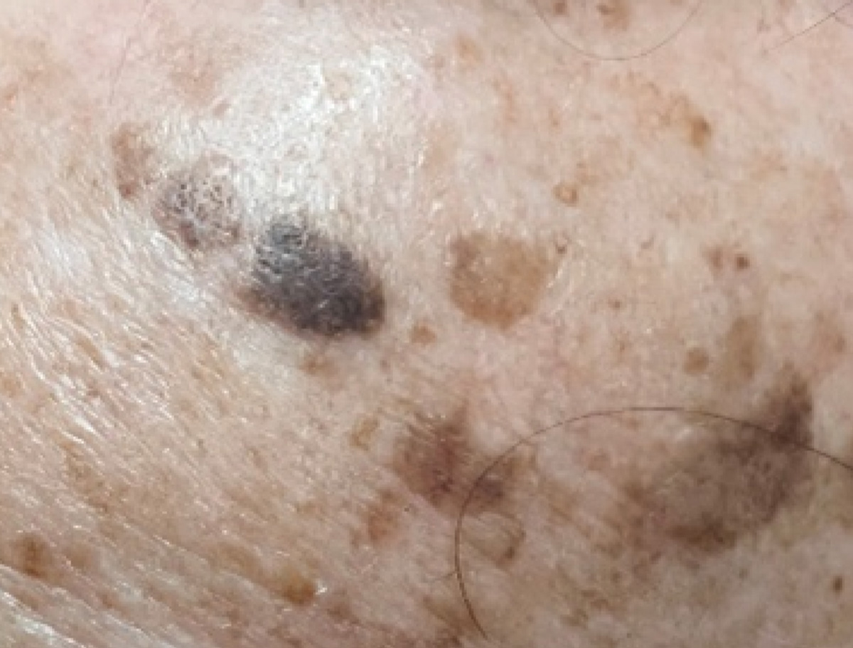
Lentigo maligna
maligna.jpg) |
| Photo: Rodney Bendure, OD, Jackie Burress, OD. Click image to enlarge. |
Believed to be caused by sun exposure, these pre-malignant lesions tend to be flat brown-to-black macules occurring in older individuals, with a median age of occurrence at 65. Their appearance has been described as looking like a stain on the skin. Slow growth and irregular borders are typical. Nodular thickening and variations in color suggest malignant transformation.
Suggested reading:
Don't Be Stumped by These Lumps and Bumps
Recognize Benign vs. Malignant Eyelid Tumors and Lesions
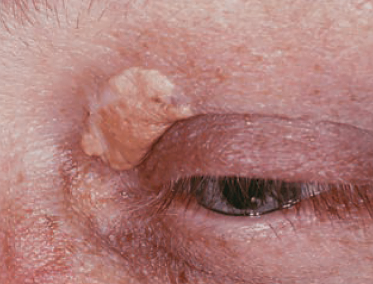
Seborrheic keratosis
 |
| Photos: Rodney Bendure, OD, Jackie Burress, OD. Click image to enlarge. |
 |
| Click image to enlarge. |
Seborrheic keratoses appear as elevated, pigmented, crusty, greasy, stuck-on plaques. While benign, a sudden increase in number or size could indicate a systemic malignancy. They develop from intradermal proliferation of basal cells within the epidermis.
Suggested reading:
Don't Be Stumped by These Lumps and Bumps
Recognize Benign vs. Malignant Eyelid Tumors and Lesions
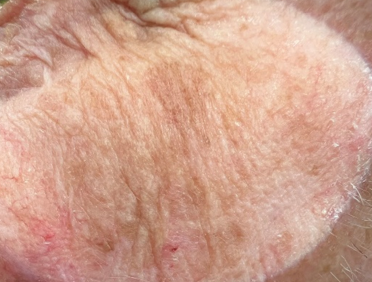
Solar lentigo
 |
| Photo: Rodney Bendure, OD, Jackie Burress, OD. Click image to enlarge. |
Solar lentigos are evenly hyperpigmented macules, while precancerous lesions have variable pigment and more irregular borders.
Suggested reading:
Don't Be Stumped by These Lumps and Bumps
Recognize Benign vs. Malignant Eyelid Tumors and Lesions
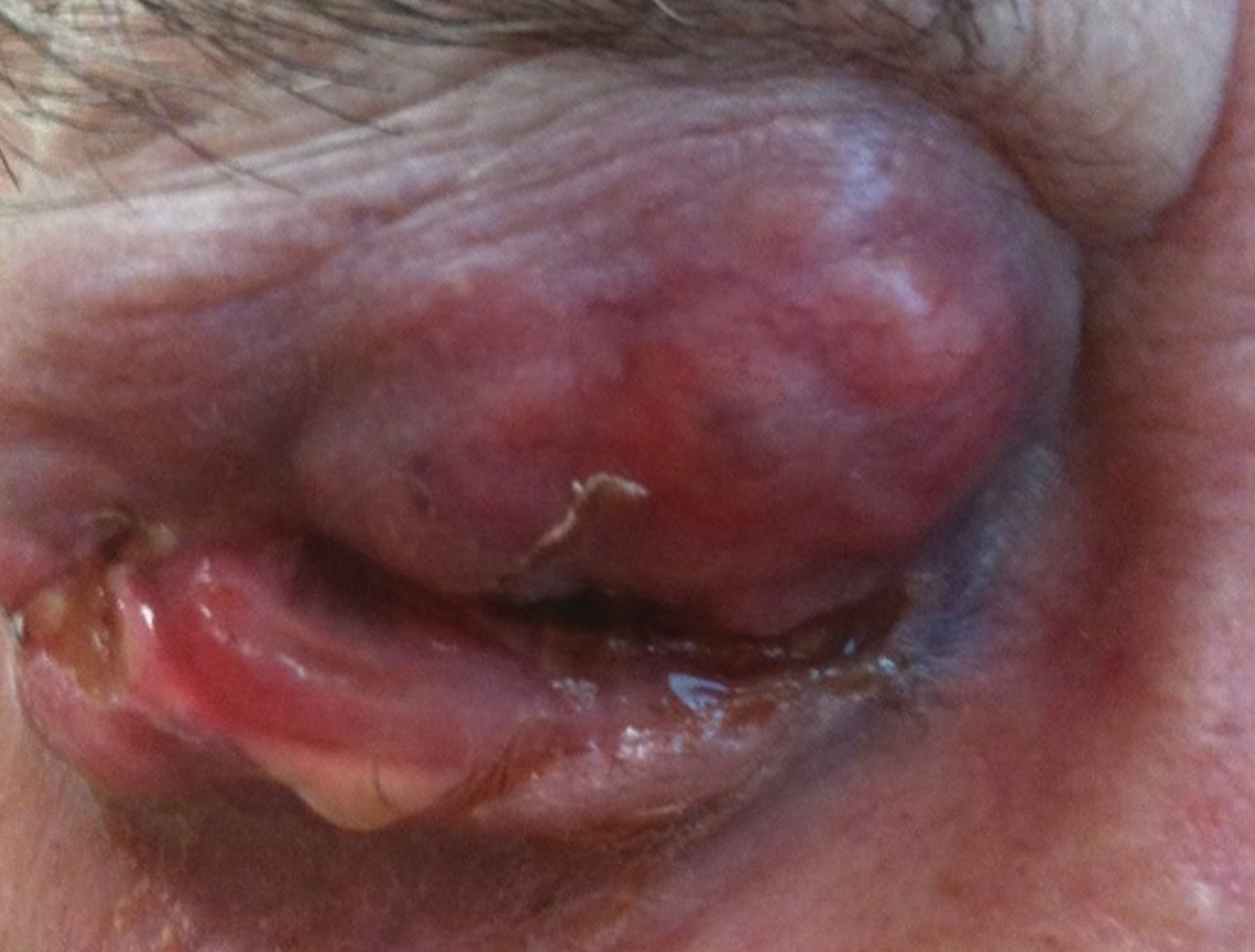
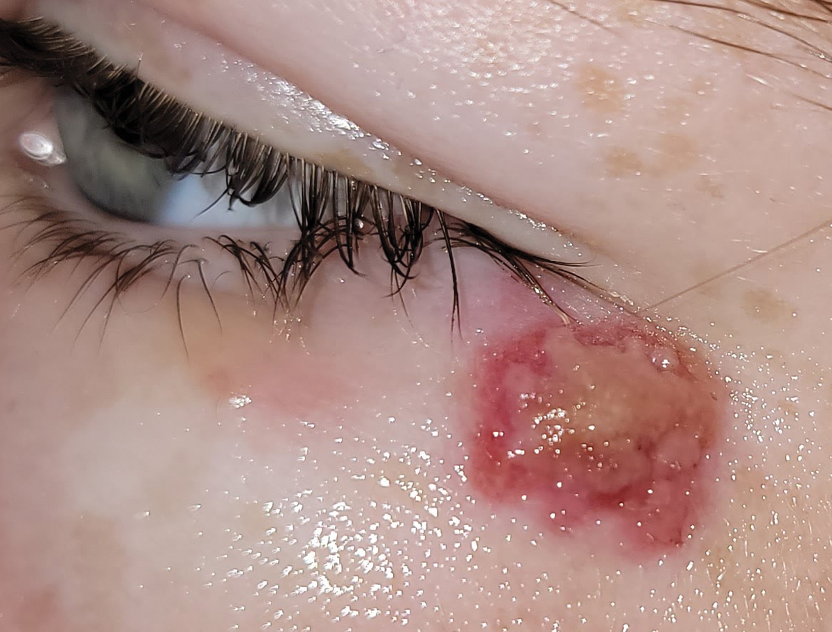
Sebaceous gland carcinoma
 |
|
Photo: Rodney Bendure, OD, Jackie Burress, OD. Click image to enlarge. |
Sebaceous gland carcinoma—a rare, slow-growing tumor with predilection for the upper lid—can be mistaken for internal hordeola.
Suggested reading:
Don't Be Stumped by These Lumps and Bumps
Recognize Benign vs. Malignant Eyelid Tumors and Lesions
Sebaceous cyst
 |
| Photos: Rodney Bendure, OD, Jackie Burress, OD. Click image to enlarge. |
 |
| Click image to enlarge. |
Sebaceous cysts, caused by blocked pilosebaceous follicles containing sebum, are more commonly found on lid margin structures such as the gland of Zeis (top) but may rarely also occur on the inner canthus, including the caruncle (bottom).
Suggested reading:
Don't Be Stumped by These Lumps and Bumps
Recognize Benign vs. Malignant Eyelid Tumors and Lesions
Blepharitis
 |
| Photos: University of Iowa. Click image to enlarge. |
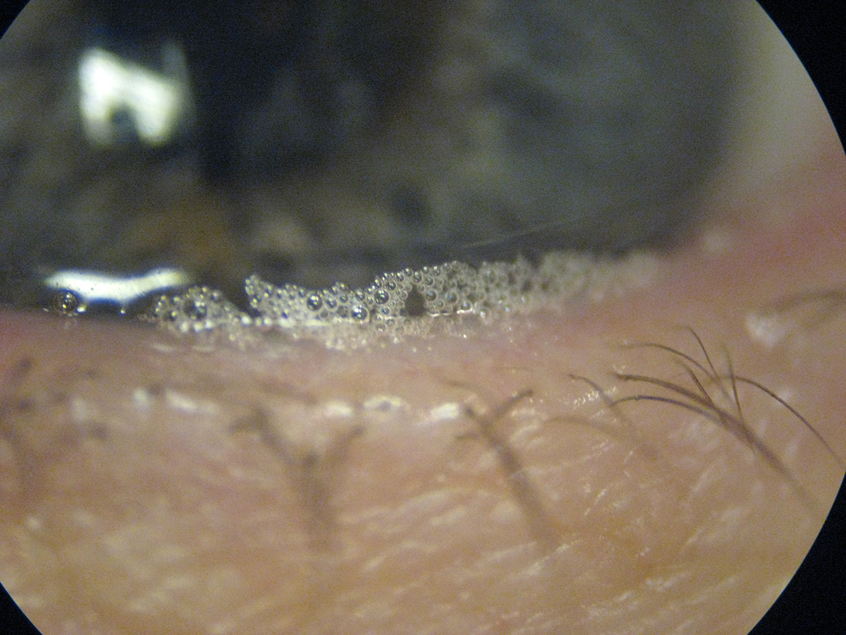 |
| Click image to enlarge. |
 |
| Click image to enlarge. |
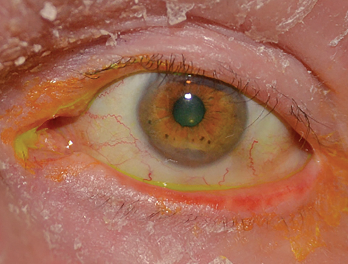
In posterior blepharitis (top), thick meibum secretions may been seen upon gland expression. Poor quality meibum in such patients can also lead to saponification of the lipid layer of the tear film. Blepharitis is part of the spectrum of ocular rosacea.
Suggested reading:
Anterior Blepharitis: The Front Line of OSD
Caruncle squamous papilloma
 |
| Photo: Rodney Bendure, OD, Jackie Burress, OD. Click image to enlarge. |
These lesions are benign and usually asymptomatic. Of all lesions involving the caruncle, papillomas account for roughly 25%. These appear as a frond-like growth with fine vascular tufts, more commonly described as resembling cauliflower. There is a strong association between conjunctival papillomas and the human papilloma virus, largely types 6 and 11.
Suggested reading:
Don't Be Stumped by These Lumps and Bumps
Recognize Benign vs. Malignant Eyelid Tumors and Lesions
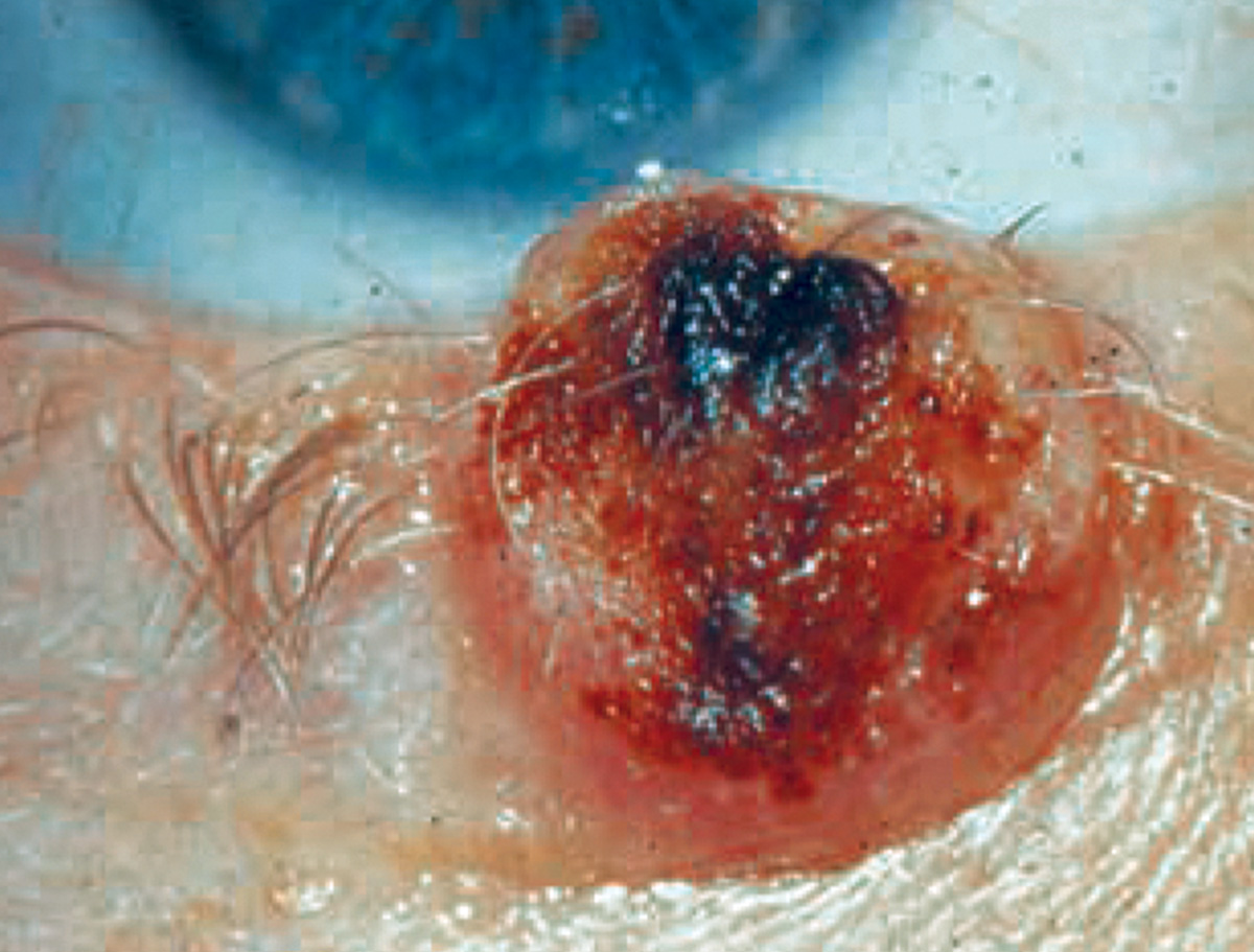
Melanoma of the punctum
 |
| Photo: Rodney Bendure, OD, Jackie Burress, OD. Click image to enlarge. |
Melanomas of the eyelids can present variably as a darkly pigmented (as shown) to an amelanotic nodule. These malignancies grow rapidly with notable bleeding and ulceration. These dangerous lesions often spread despite aggressive excision efforts with controlled surgical margins. Patients will need close follow-up care due to risk of metastasis.
Suggested reading:
Recognize Benign vs. Malignant Eyelid Tumors and Lesions
A Close Look at Common Lid Lesions
Keratoacanthoma
 |
| Photo: Rodney Bendure, OD, Jackie Burress, OD. Click image to enlarge. |
These pre-malignant lesions present as a dome-shaped nodule with a keratin-filled core on the sun-exposed skin of individuals over the age of 50. These usually develop rapidly over weeks, only to spontaneously regress after a few months. However, if the lesion persists, it may be removed by Mohs surgery.
Suggested reading:
Don't Be Stumped by These Lumps and Bumps
Recognize Benign vs. Malignant Eyelid Tumors and Lesions
Ecthyma contagiosum
 |
| Photo: University of Iowa. Click image to enlarge. |
Ecthyma contagiosum, caused by a parapoxvirus. Spread from sheep/goats to humans. Differential is other viruses, such as HSV/ HZV, and tumor.
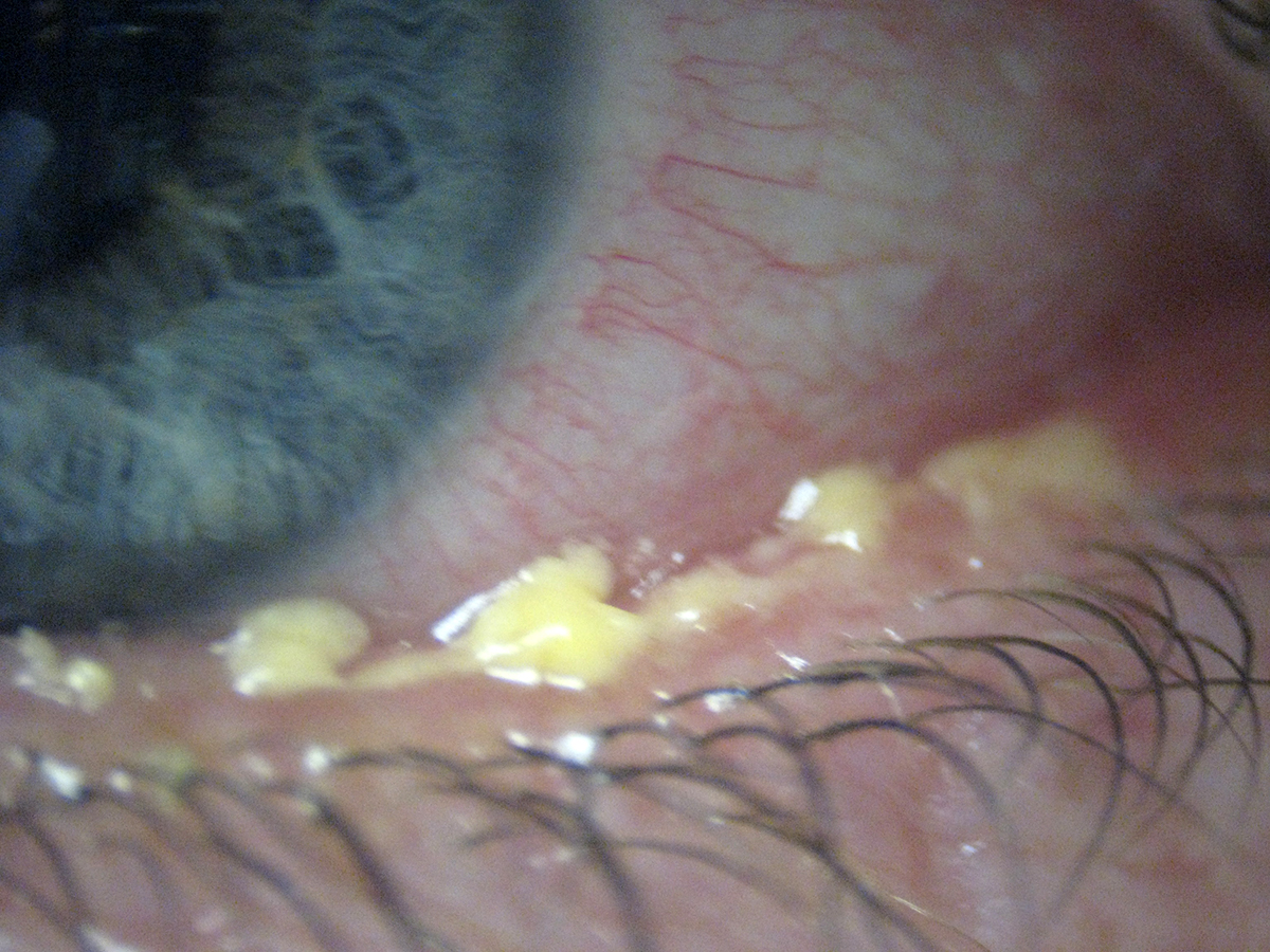
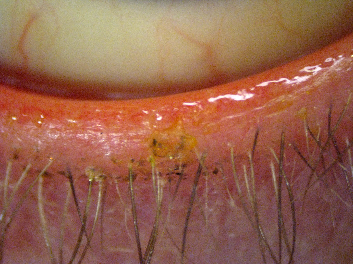
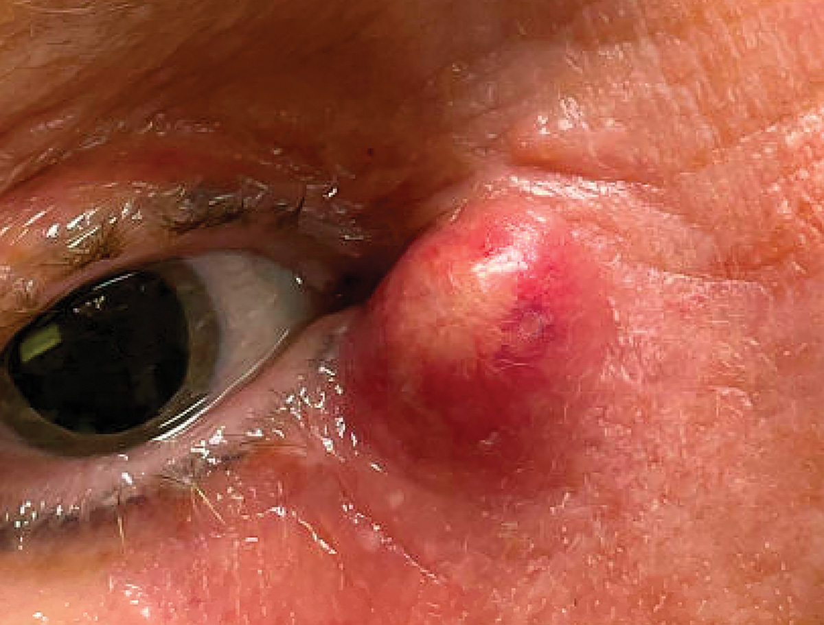
Chalazion & hordeolum
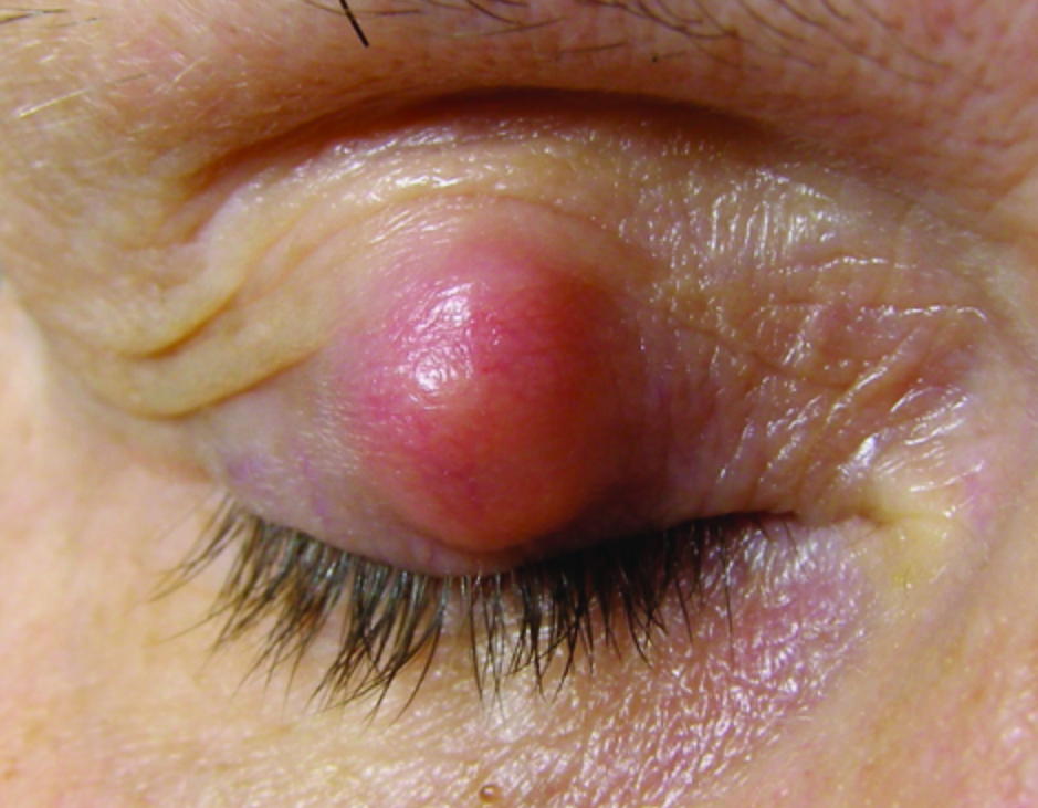 |
| Photo: Nate Lighthizer, OD. Click image to enlarge. |
 |
| Photo: Greg Caldwell, OD. Click image to enlarge. |
Chalazion (top) and hordeolum (bottom) are often confused for each other. Chalazia may present with acute inflammation but more often are non-tender. External hordeola involve lash follicles, while internal forms are often due to bacterial infection of a meibomian gland.
Suggested reading:
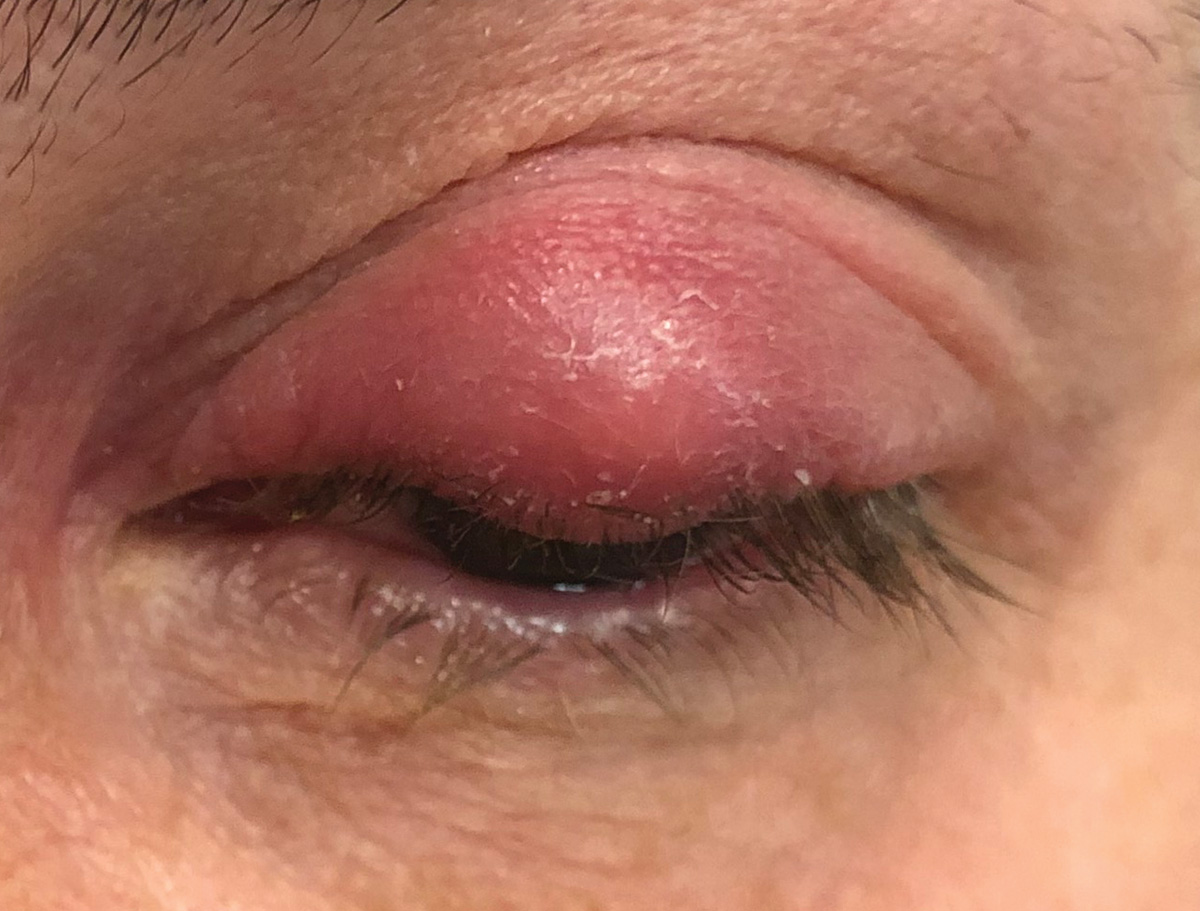
Ocular rosacea
 |
| Photo: University of Iowa. Click image to enlarge. |
Ocular rosacea, an oft-overlooked serious chronic inflammatory condition of the lid margins, causing blurred vision and pain.
Suggested reading:
Seeing Red: How Ocular Rosacea Impacts the Cornea
Differentiating Problems of the Eyelids and Ocular Adnexa
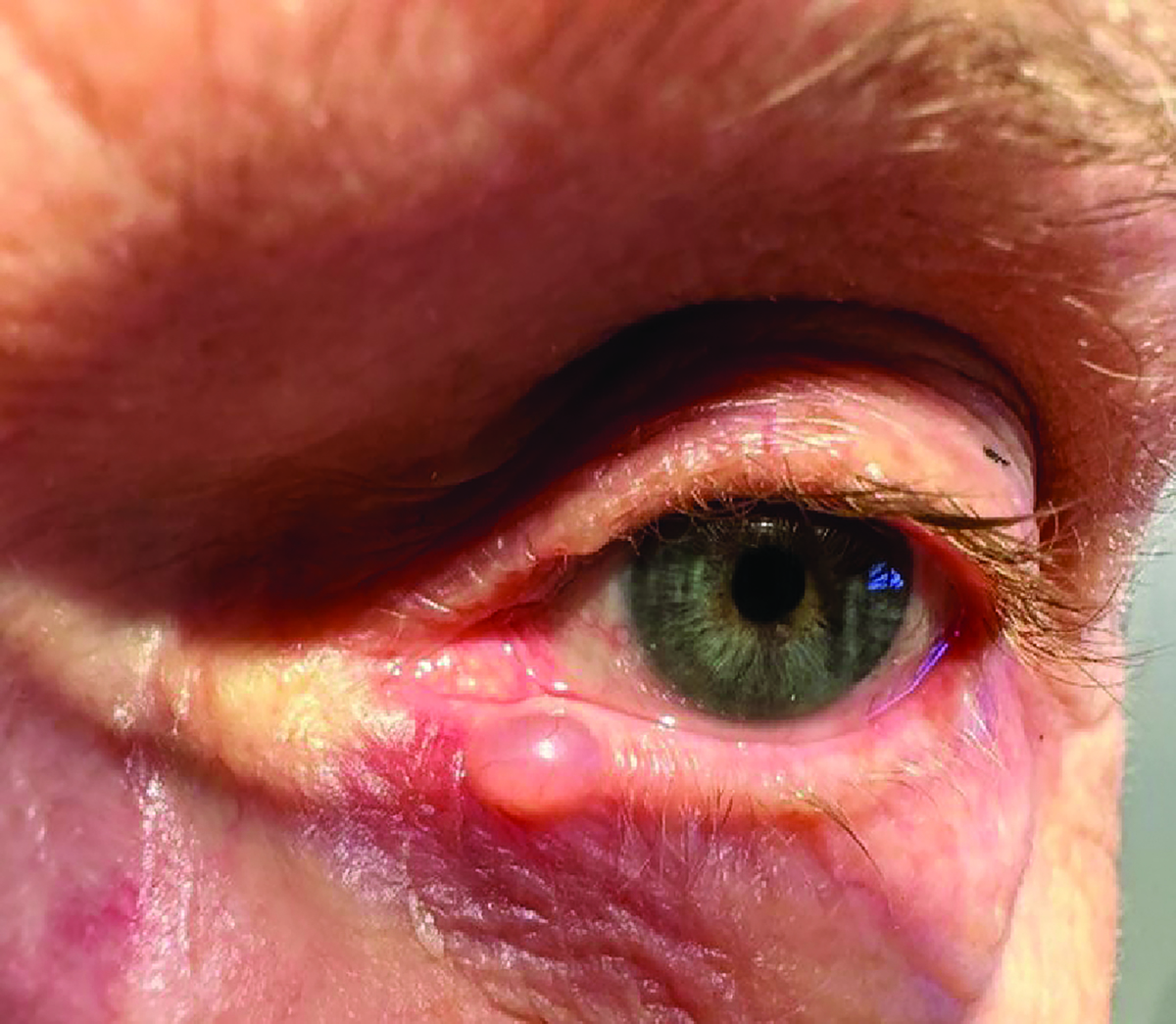
Eccrine cyst
 |
| Photo: Rodney Bendure, OD, Jackie Burress, OD. Click image to enlarge. |
Eccrine cysts are small, smooth, translucent nodules associated with sweat glands. They often grow in size during hot, humid weather.
Suggested reading:
Don't Be Stumped by These Lumps and Bumps
Recognize Benign vs. Malignant Eyelid Tumors and Lesions
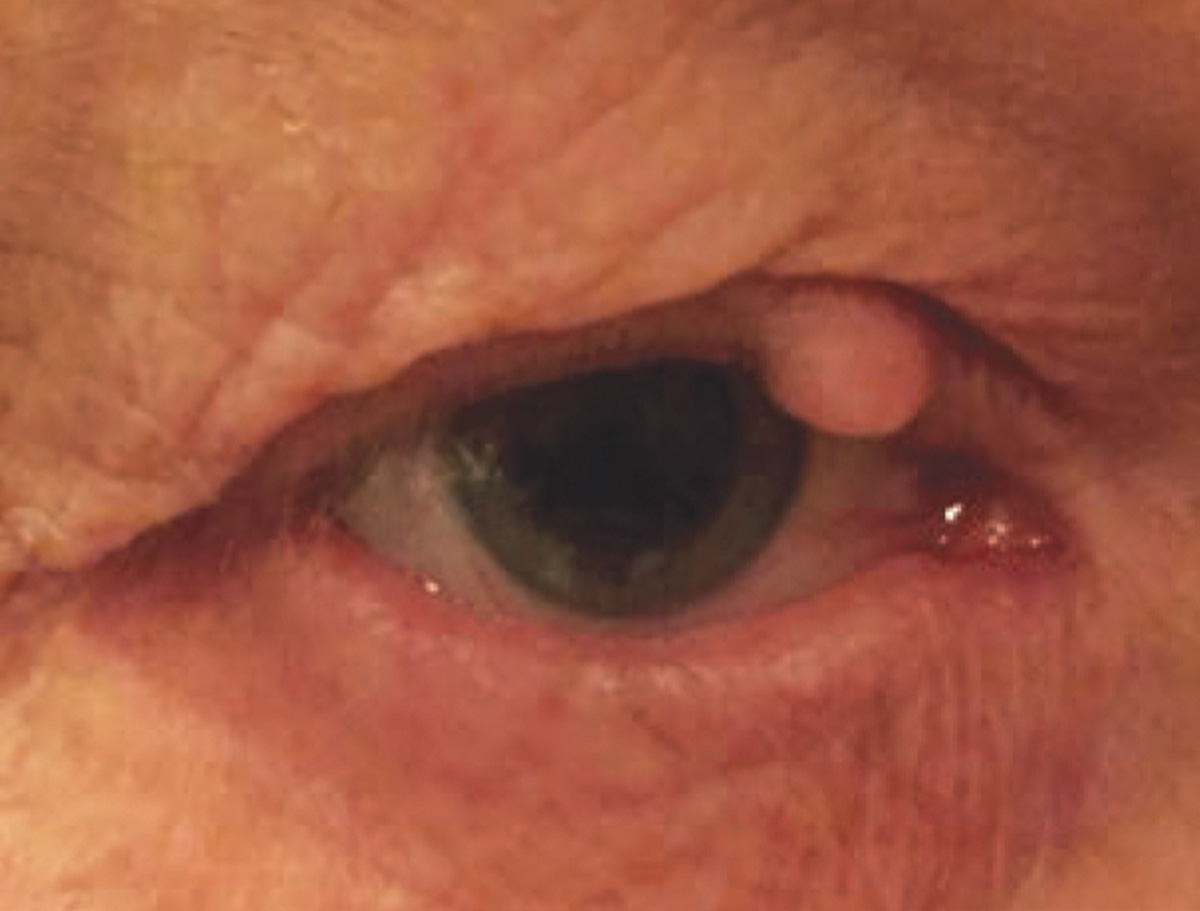
Epidermal inclusion cysts
 |
| Photo: Rodney Bendure, OD, Jackie Burress, OD. Click image to enlarge. |
These arise from entrapment (usually traumatic) of epidermal tissue within the dermis. They appear as discrete white or light yellow, firm, solid, slow-growing cysts. Treatment is by excision, though the entire cyst wall must be removed to prevent recurrence.
Suggested reading:
Don't Be Stumped by These Lumps and Bumps
Recognize Benign vs. Malignant Eyelid Tumors and Lesions
Verruca vulgaris
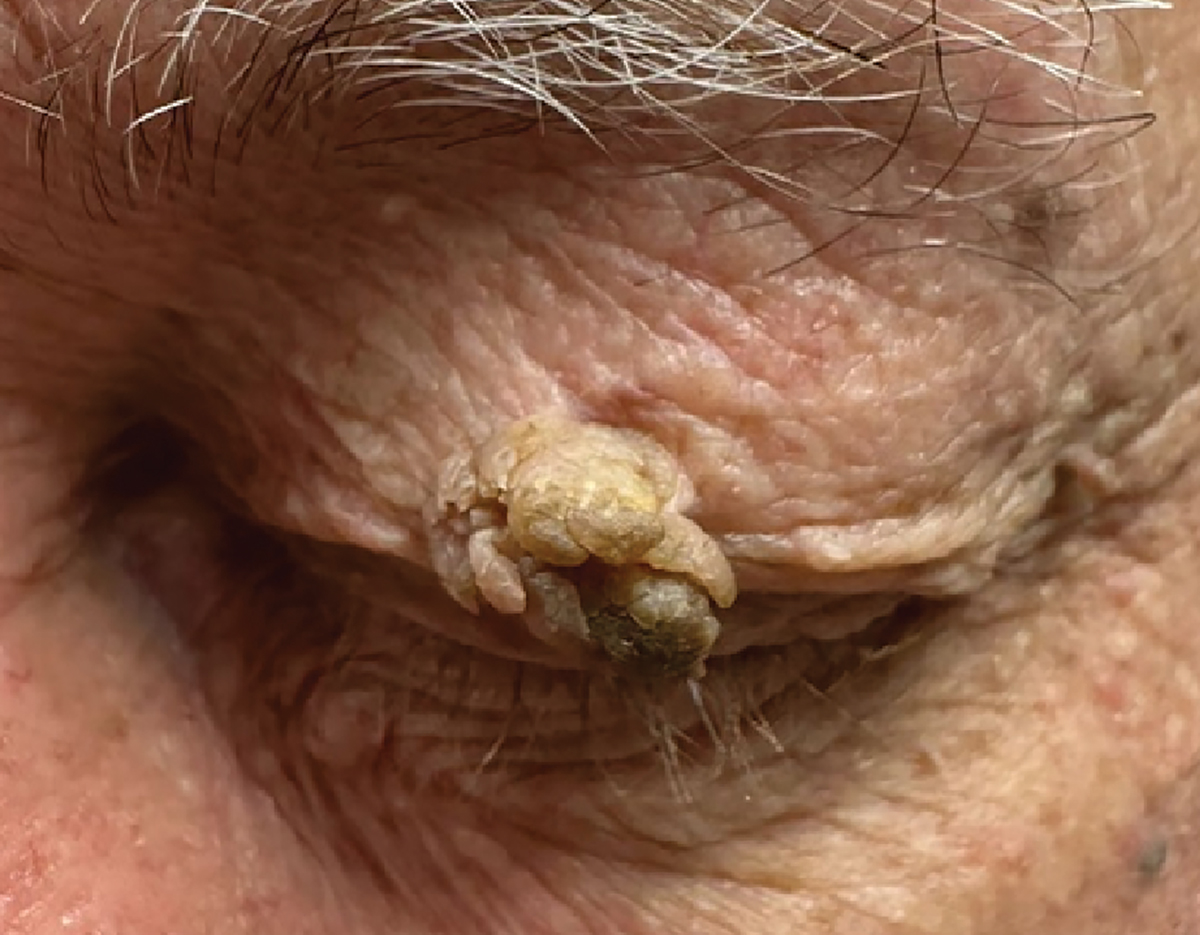 |
| Photo: Rodney Bendure, OD, Jackie Burress, OD. Click image to enlarge. |
Also known as a viral wart, this is an epidermal growth caused by the human papilloma virus, typically types VI or XI. Two forms exist: filiform, which are also called digitate because they project in a finger-like fashion from their base, and plana, which are flat. Observation is often adequate, as these lesions tend to eventually outgrow their blood supply and spontaneously involute, but may be removed by excision, cryotherapy or chemical cautery if eye irritation ensues or for cosmetic reasons.
Suggested reading:
Recognize Benign vs. Malignant Eyelid Tumors and Lesions
Differentiating Problems of the Eyelids and Ocular Adnexa
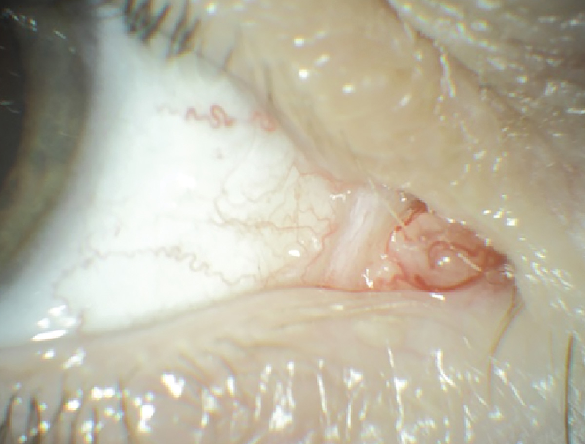
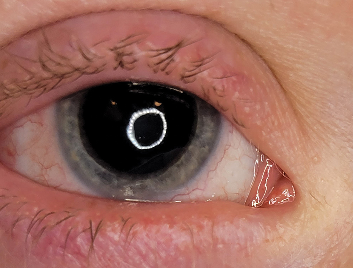
Dacryocystitis
 |
| Photo: Alison Bozung, OD. Click image to enlarge. |
Dacryocystitis is an infection of the lacrimal sac, commonly attributed to an acquired nasolacrimal duct obstruction.caption
Suggested reading:
Managing Dacryoceles And Dacryocystitis
Dacryoadenitis
 |
| Photos: Alison Bozung, OD. Click image to enlarge. |
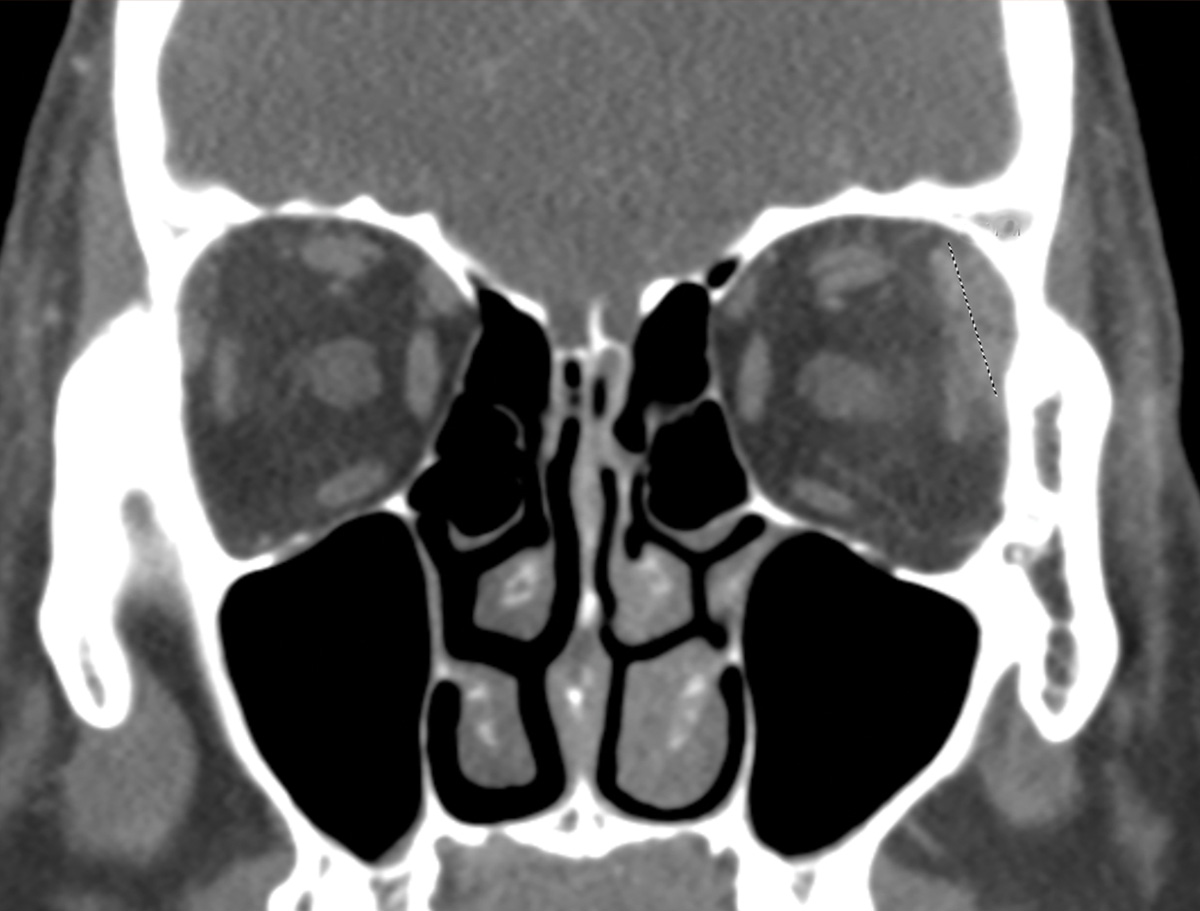 |
| Click image to enlarge. |
Dacryoadenitis refers to inflammation of the lacrimal gland. It may be caused by an autoimmune condition (i.e., sarcoidosis, Sjögren’s syndrome, IgG4 disease), an infection (i.e., Epstein-Barr Virus, adenoviral, bacterial) or can be idiopathic.
Suggested reading:
Keeping an Eye Out for Lacrimal Gland Abnormalities
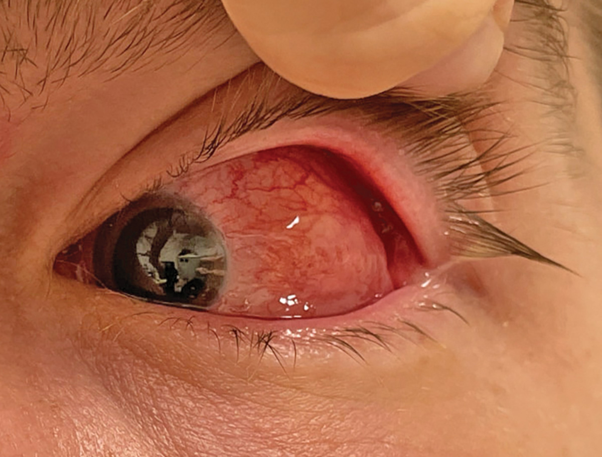
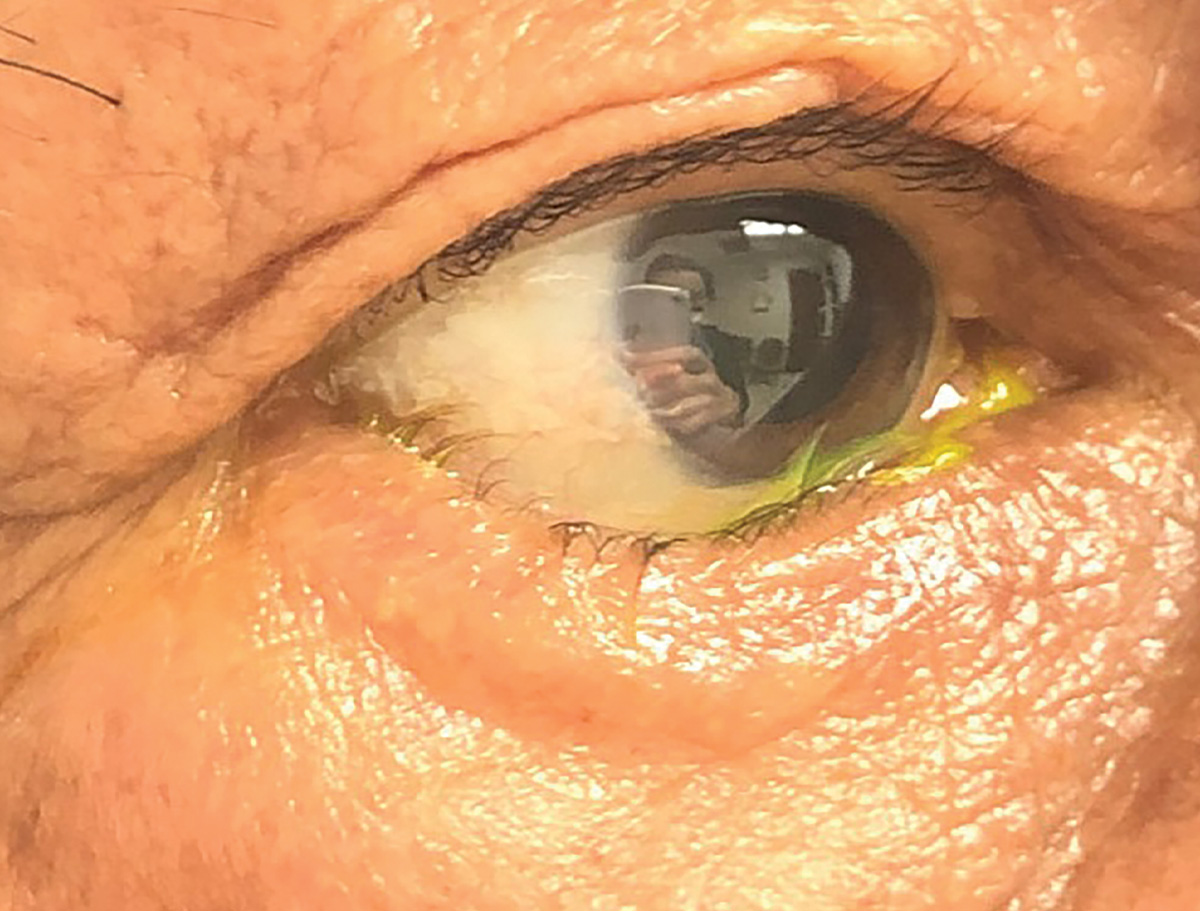
Cicatricial ectropion
 |
| Photo: Alison Bozung, OD. Click image to enlarge. |
Cicatricial ectropion, secondary here to cutaneous T-cell lymphoma, may lead to significant ocular surface exposure.
Suggested reading:
A Game Plan for Managing Eyelid Lesions and Related Conditions
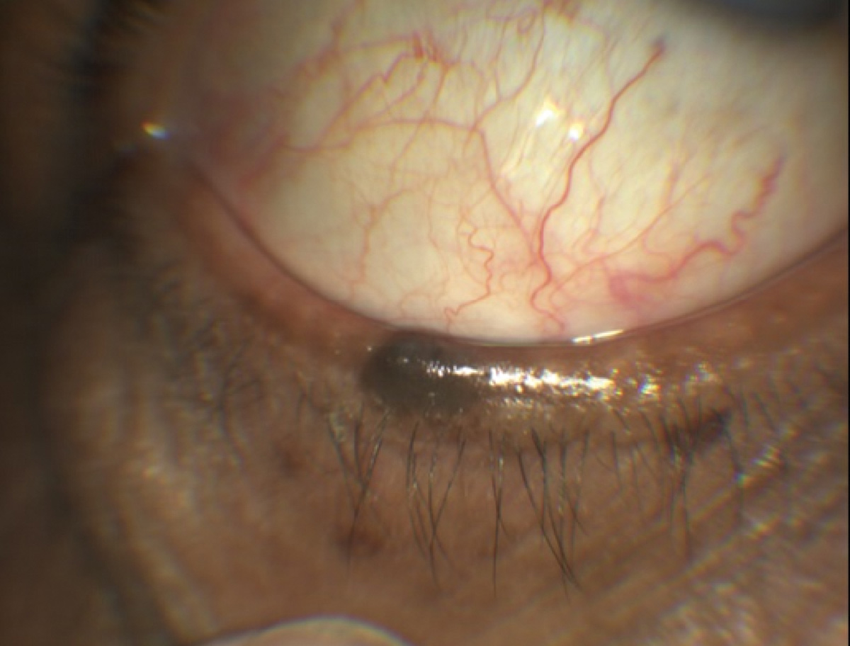
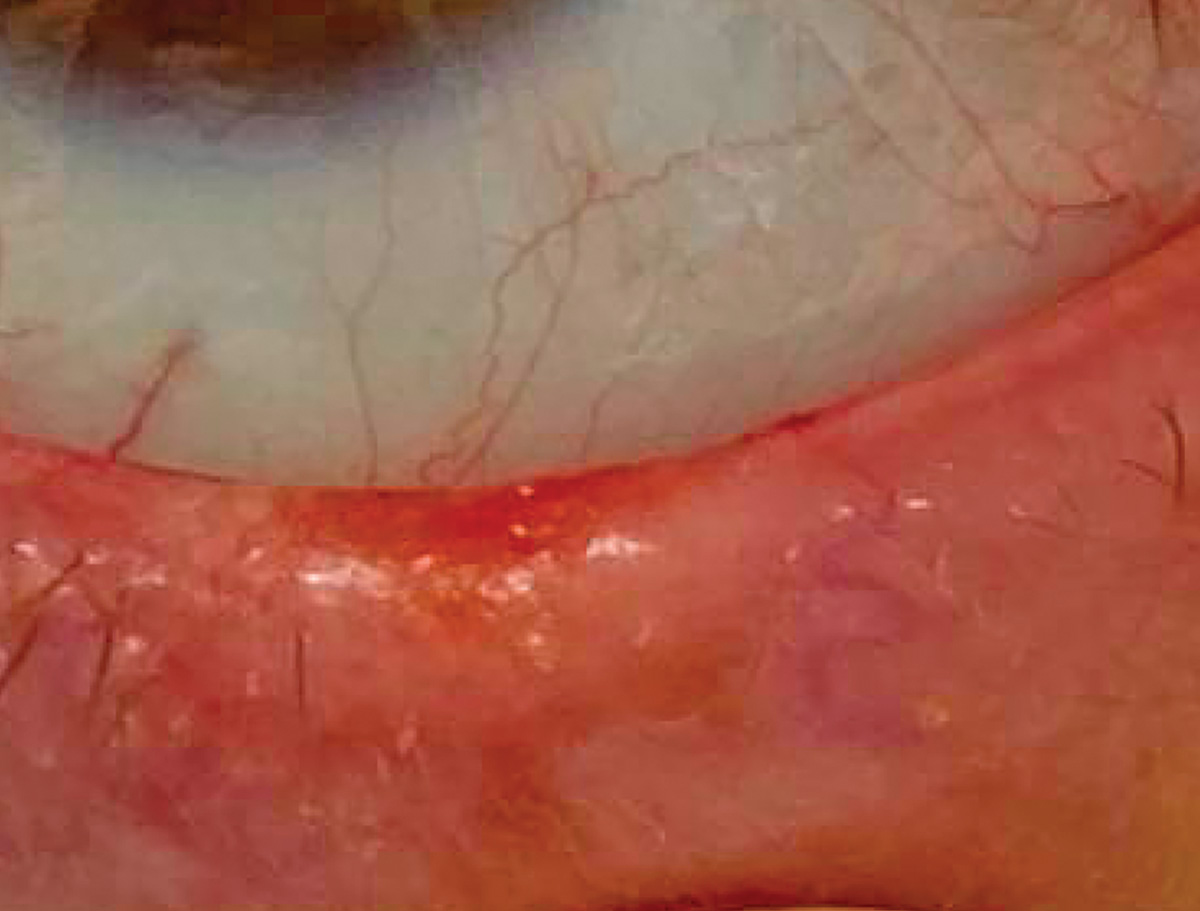
Entropion
 |
| Photo: Alison Bozung, OD. Click image to enlarge. |
Spastic entropion is intermittent and may only be evident after squeezing the eyelids closed, as in this patient.
Suggested reading:
A Game Plan for Managing Eyelid Lesions and Related Conditions
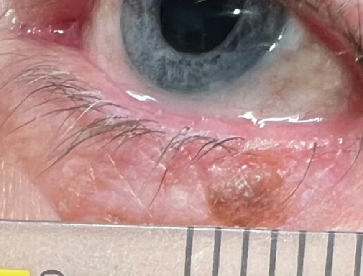
Basal cell carcinoma
 |
| Photo: Alison Bozung, OD. Click image to enlarge. |
Basal cell carcinoma, the most common skin cancer, often has vascular pearly borders. Always assess for lid madarosis.
Suggested reading:
A Game Plan for Managing Eyelid Lesions and Related Conditions
