Photo Atlas of Ocular DiseaseReview of Optometry has created the following new resource—a photo atlas of ocular diseases across the entire spectrum of eye care. In addition to over 200 high-quality photos, you'll find clinical pearls for each condition and links to relevant articles that provide detailed guidance. An index of the entire atlas can be found here or you can check out the conditions featured in their respective categories: |
Sight, a remarkable sensation, involves a complex journey of light through the tear film, anterior chamber, pupil, crystalline lens and vitreous, followed by processing in the retina. While the interplay of these components is crucial for vision, the optic nerve holds an especially pivotal role in transmitting visual messages to the brain.
Often associated with glaucoma, the problems of the optic nerve encompass a broader spectrum of conditions beyond this common perception. Numerous conditions—ranging from congenital diseases like coloboma to toxic neuropathies, neurodegenerative diseases and ocular systemic disorders—affect the optic nerve’s structure and function.
The physical examination of the optic nerve, which has always been a critical part of an eye exam, has its limitations, primarily as we are confined to the observable optic nerve head. However, there are several functional and structural tests, including various photographic techniques and OCT, that can aid us in assessing the function of the optic nerve and in differential disease diagnosis.
Feel free to click the condition that interests you to jump to it, or scroll to look through all the conditions.
Idiopathic intracranial hypertension
Leber's hereditary optic neuropathy
Anterior ischemic optic neuropathy
Neuroretinitis
 |
| Photos: Jessica Haynes, OD. Click image to enlarge. |
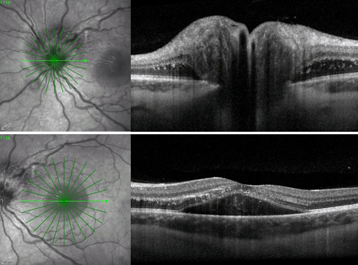 |
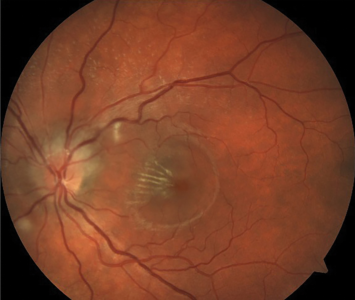
| Click image to enlarge. |
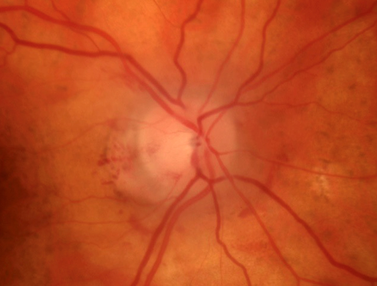
Neuroretinitis is defined as inflammation or swelling of the anterior optic nerve and the macula. It has a wide range of etiologies from infectious to idiopathic, with cat scratch disease from Bartonella being the most common cause of infectious neuroretinitis. This patient presents with swelling of the optic nerve and macula. They have a partial macular star forming as exudates radiate away from the fovea. The OCT images show corresponding swelling of the optic nerve and macular edema.
Suggested reading:
A Case of Bilateral Bartonella Henselae Neuroretinitis
Idiopathic intracranial hypertension
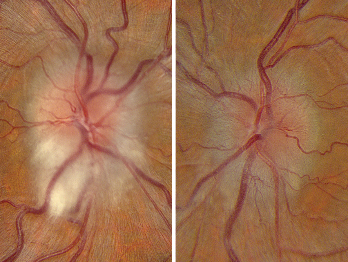 |
| Photos: Alison Bozung, OD. Click image to enlarge. |
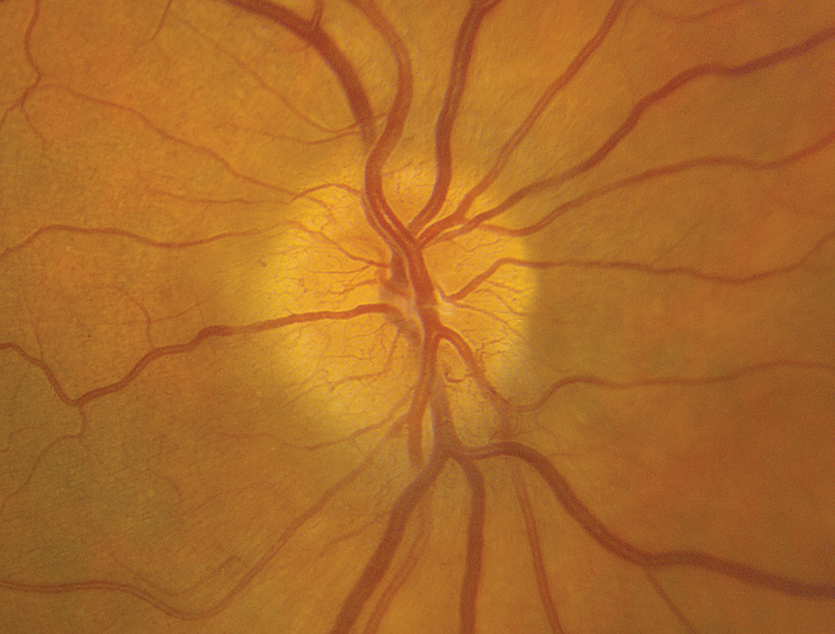
IIH typically presents with bilateral disc edema, transient visual obscurations, headaches, and pulsatile tinnitus. This patient also had myelinated nerve fiber layer in the right eye.
Suggested reading:
The Swollen Optic Disc: Is this an Emergency?
IIH Missed or Prematurely Diagnosed in Literature by Almost 20%
Atrophic Optic Discs in IIH Patients Can Experience Recurrent Swelling
Leber’s hereditary optic neuropathy
 |
| Photos: Alison Bozung, OD. Click image to enlarge. |
Leber’s begins unilaterally but rapidly involves the fellow eye. Patients develop painless central scotomas.
Suggested reading:
A Detailed Look at Optic Nerve Anomalies
Are You Missing These Optic Nerve Disorders?
RNFL Thinning Patterns Found In Leber’s
Six Questions About the Role of OCT in Neuro Evaluations
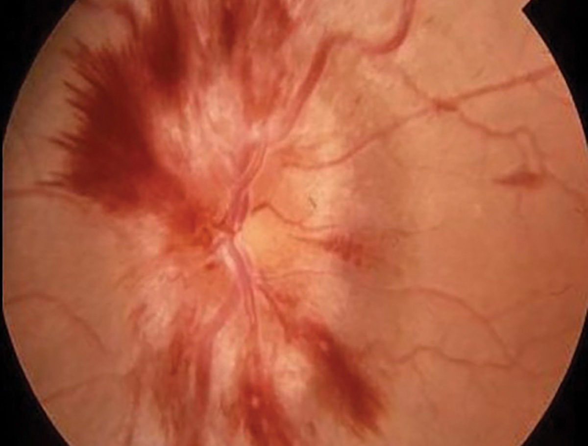
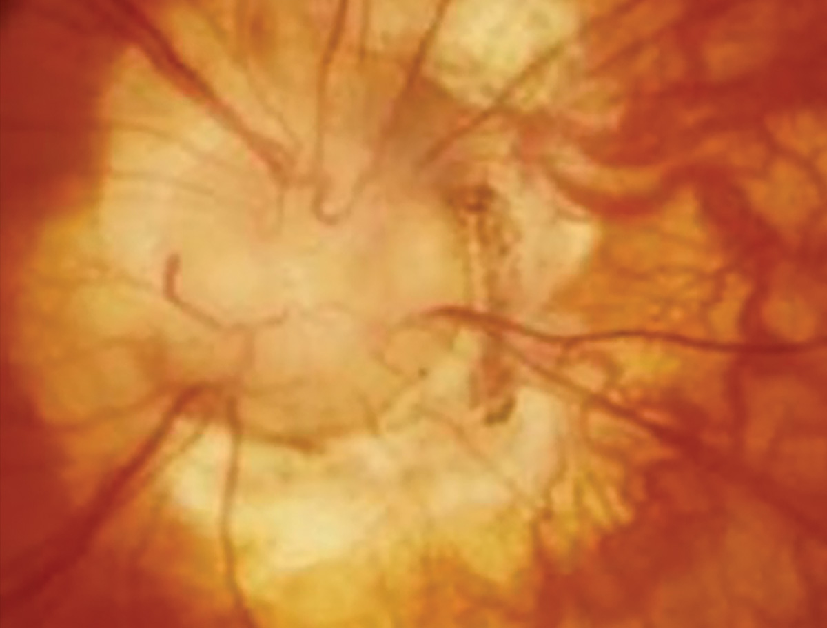
Anterior ischemic optic neuropathy
 |
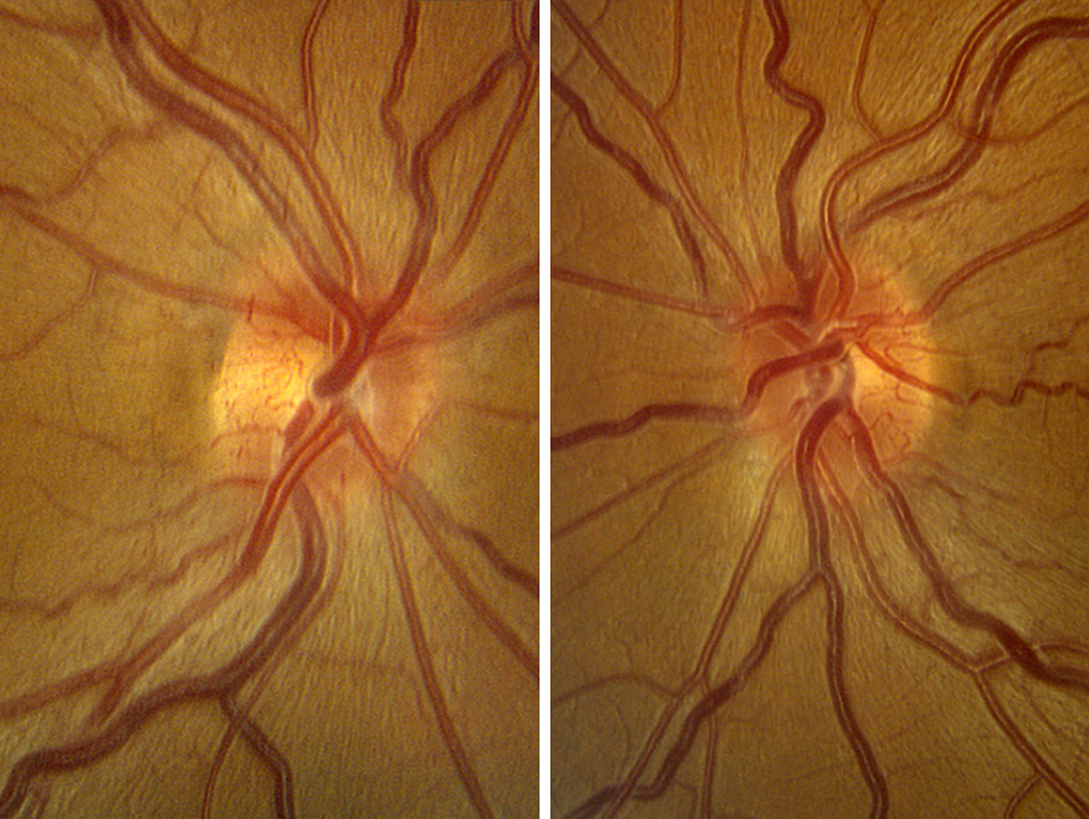
| Photos: Alison Bozung, OD. Click image to enlarge. |
 |
| Click image to enlarge. |
The first photo is giant cell arteritis with arteritic anterior ischemic optic neuropathy (AAION). The non-arteritic form of AION (NAION, second photo) is most common in individuals with a history of cardiovascular disease, sleep apnea, and/or minimal optic nerve cupping. The NAION image shows circumferential disc edema, but it is most prominent inferiorly.
Suggested reading:
How to Triage Non-Traumatic Ocular Emergencies
A Detailed Look at Optic Nerve Anomalies
Optic disc drusen
 |
| Photos: Jessica Haynes, OD. Click image to enlarge. |
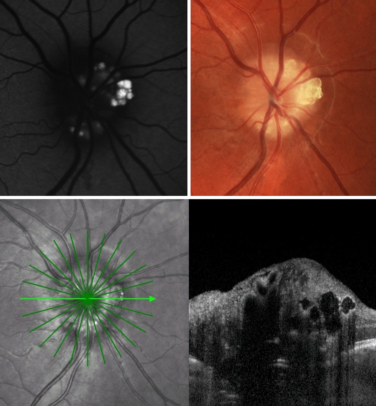
Optic disc drusen are proteinaceous deposits of the optic nerve that become calcified with age. They can be buried or superficial. More superficial and calcified disc drusen are hyperautofluoresecent, as seen in the top left FAF image. They may be visible clinically as seen on the top right photo. On OCT cross section scan (bottom image), they may be visualized as internally hyporeflective lesions with a patchy/bumpy hyper-reflective border. Optic disc drusen can lead to retinal nerve fiber layer atrophy and visual field defects. They also predispose patients to pathology such as NAION and peripapillary CNV. In addition, they can mimic optic disc edema and are a common reason for pseudopapilledema.
Suggested reading:
Optic Disc Drusen Pinpointed As Risk Factor of Nonarteritic AION
A Detailed Look at Optic Nerve Anomalies
Discern Optic Nerve Head Drusen from True Papilledema
Understanding ONH Dynamics in Glaucoma and Beyond
MOG optic neuropathy
 |
| Photo: Alison Bozung, OD. Click image to enlarge. |
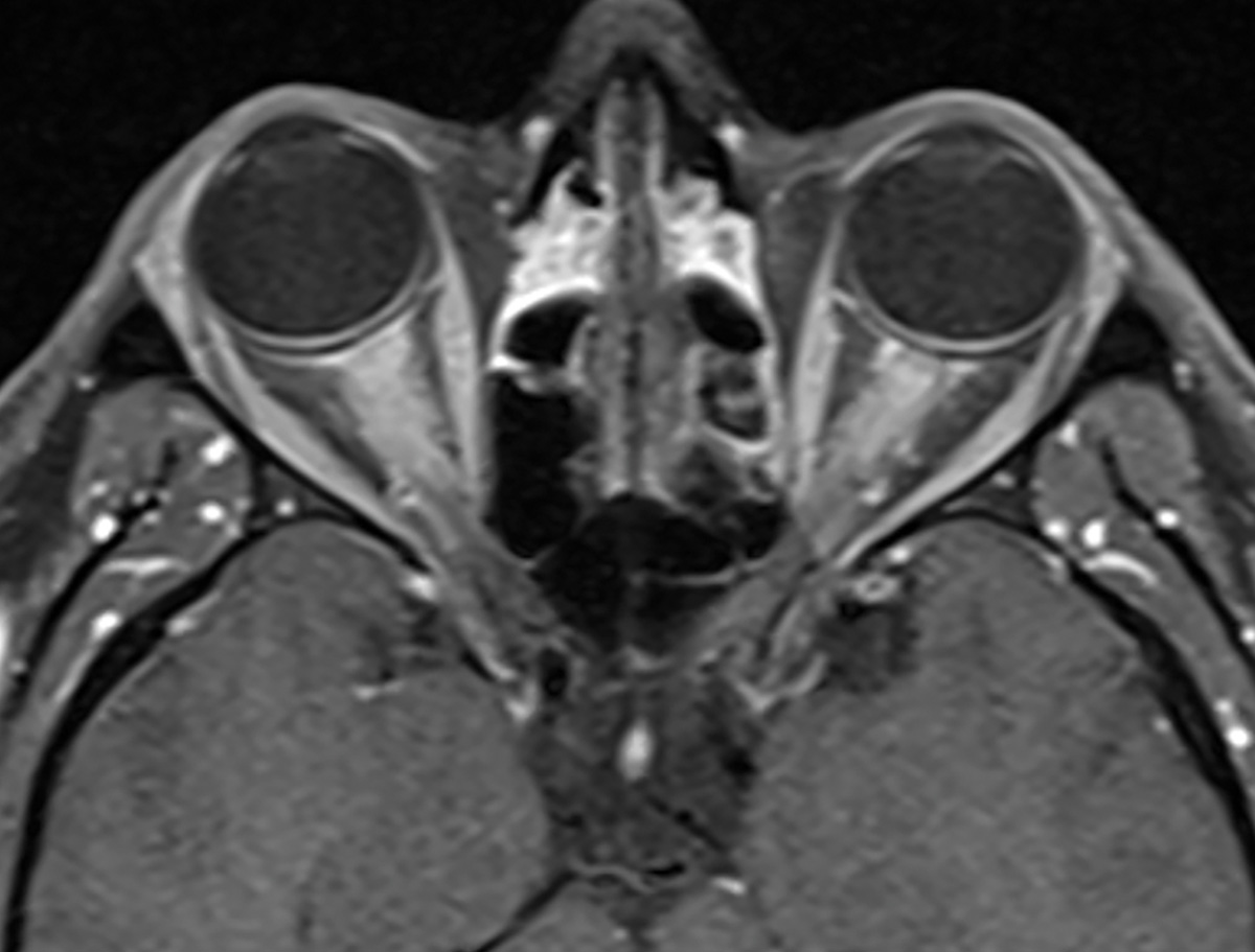
Bilateral extensive intraorbital optic nerve enhancement in myelin oligodendrocyte glycoprotein–associated optic neuropathy.
Suggested reading:
A Guide to Demyelinating Diseases of the CNS
Are You Missing These Optic Nerve Disorders?
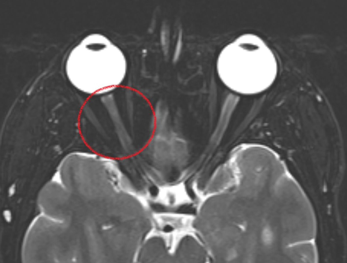
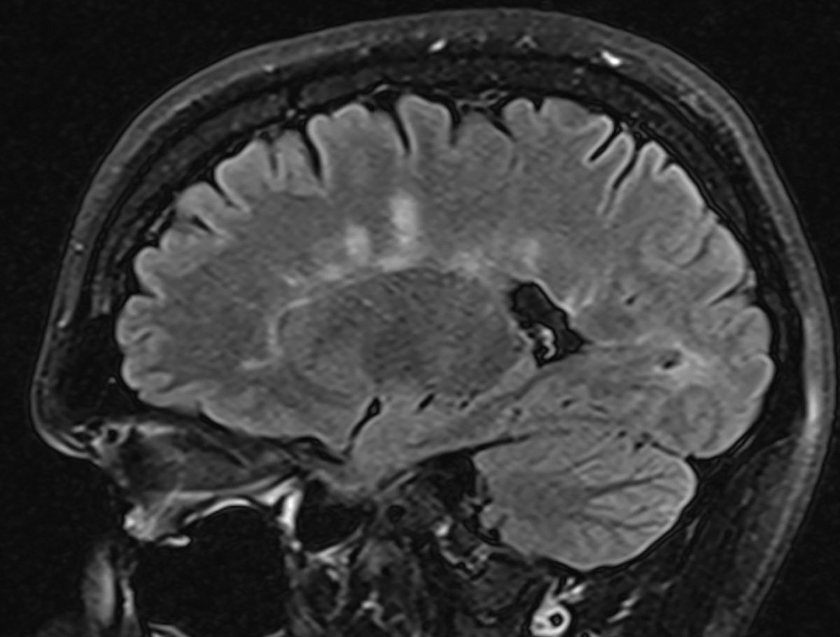
Optic neuritis
 |
| Photos: Alison Bozung, OD. Click image to enlarge. |
 |
| Click image to enlarge. |
This patient with right retrobulbar optic neuritis presented with subacute vision loss and pain on eye movement. An MRI brain revealed periventricular white matter lesions, suggestive of a demyelinating disease.
Suggested reading:
A Guide to Demyelinating Diseases of the CNS
New Biomarkers Spur Re-evaluation of Neuritis Incidence
Optic Nerve Disorders: How They Manifest and What They Mean
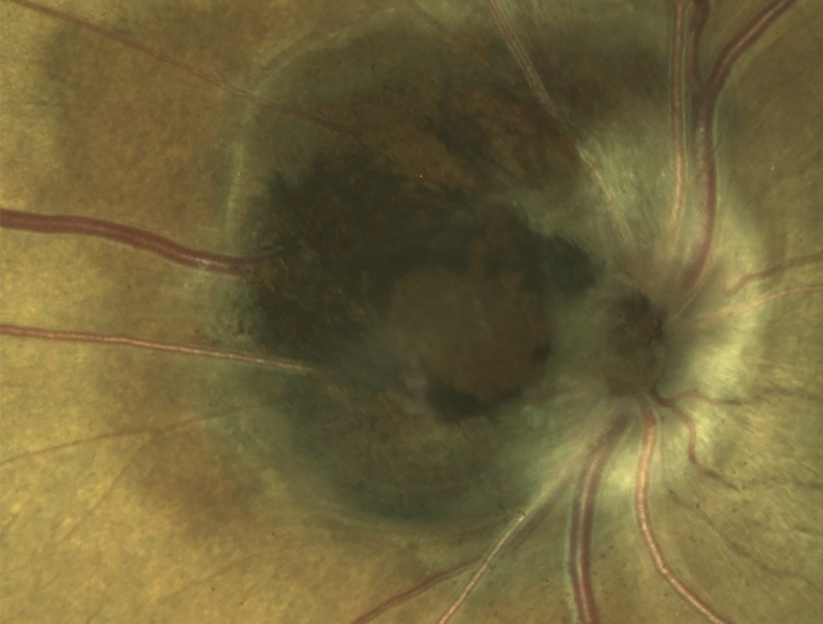
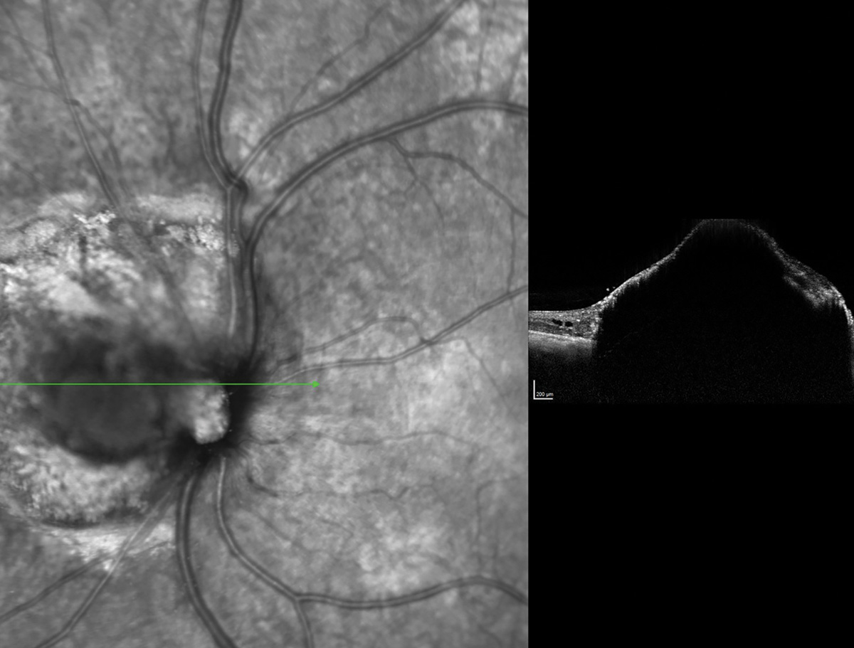
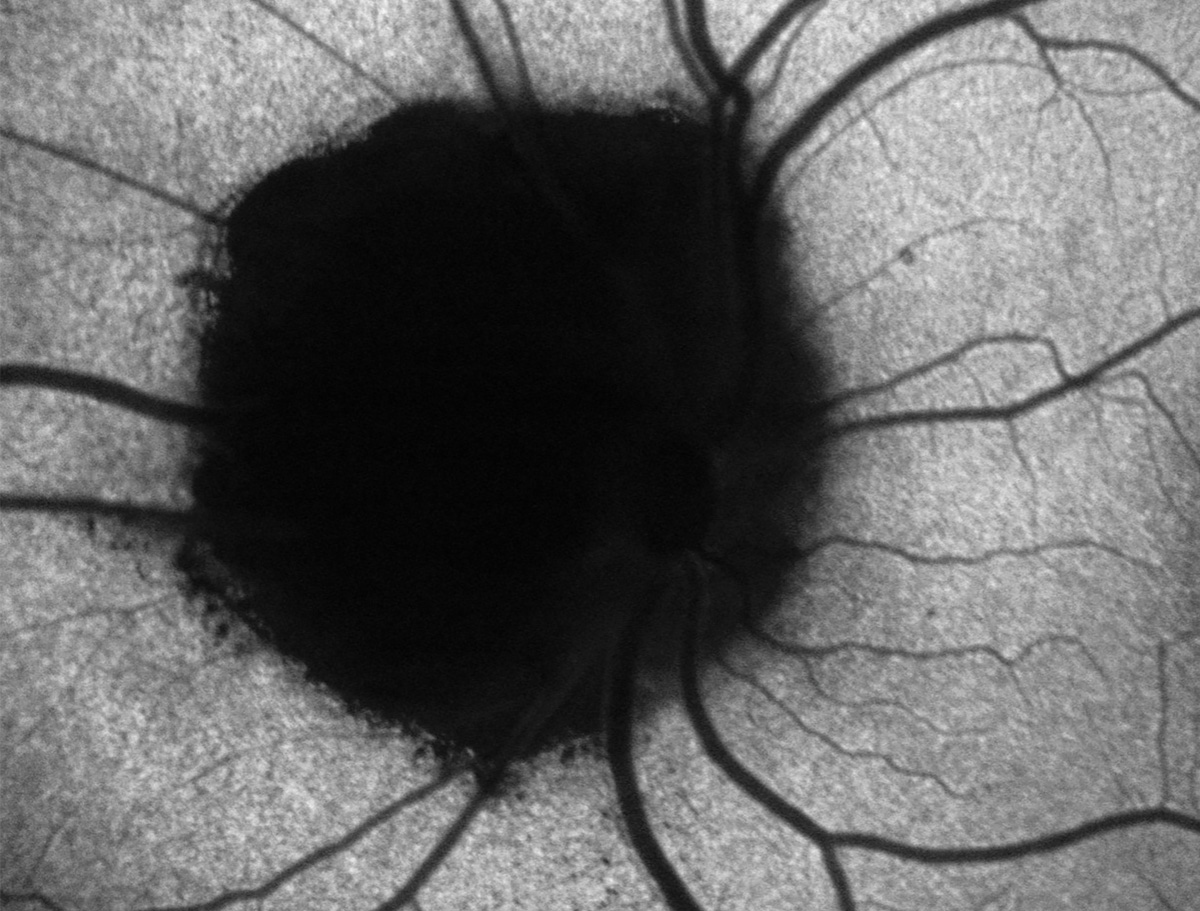
Optic nerve melanocytoma
 |
|
Photos: Mohammad Rafieetary, OD. Click image to enlarge. |
 |
| Click image to enlarge. |
 |
| Click image to enlarge. |
Optic nerve melanocytomas are rare, benign, deeply pigmented lesions. They are often distinguished by clinical appearance alone (first image), which is quite striking, but additional testing could help differentiate them from other pigmented lesions. OCT (second image) shows elevated lesion with dense superficial hyperreflectivity and posterior shadowing. On FAF (third image), the melanocytoma is hypoautofluorescent.
Suggested reading:
A Detailed Look at Optic Nerve Anomalies
Are You Missing These Optic Nerve Disorders?
Disc pallor
 |
|
Photo: Mohammad Rafieetary, OD. Click image to enlarge. |
This patient has optic atrophy and resultant disc pallor. It represents irreversible damage to the ganglion cell axons.
Suggested reading:
Are You Missing These Optic Nerve Disorders?
Shedding Light on a Pale Optic Nerve
Papilledema
 |
| Photo: Mohammad Rafieetary, OD. Click image to enlarge. |
Papilledema may lead to elevation of the neuroretinal rim, indistinct disc margins, hemorrhages and Paton’s lines.
Suggested reading:
How to Triage Non-Traumatic Ocular Emergencies
Optic Nerve Disorders: How They Manifest and What They Mean
Discern Optic Nerve Head Drusen from True Papilledema
Term “Papilledema” Often Misused in the Literature
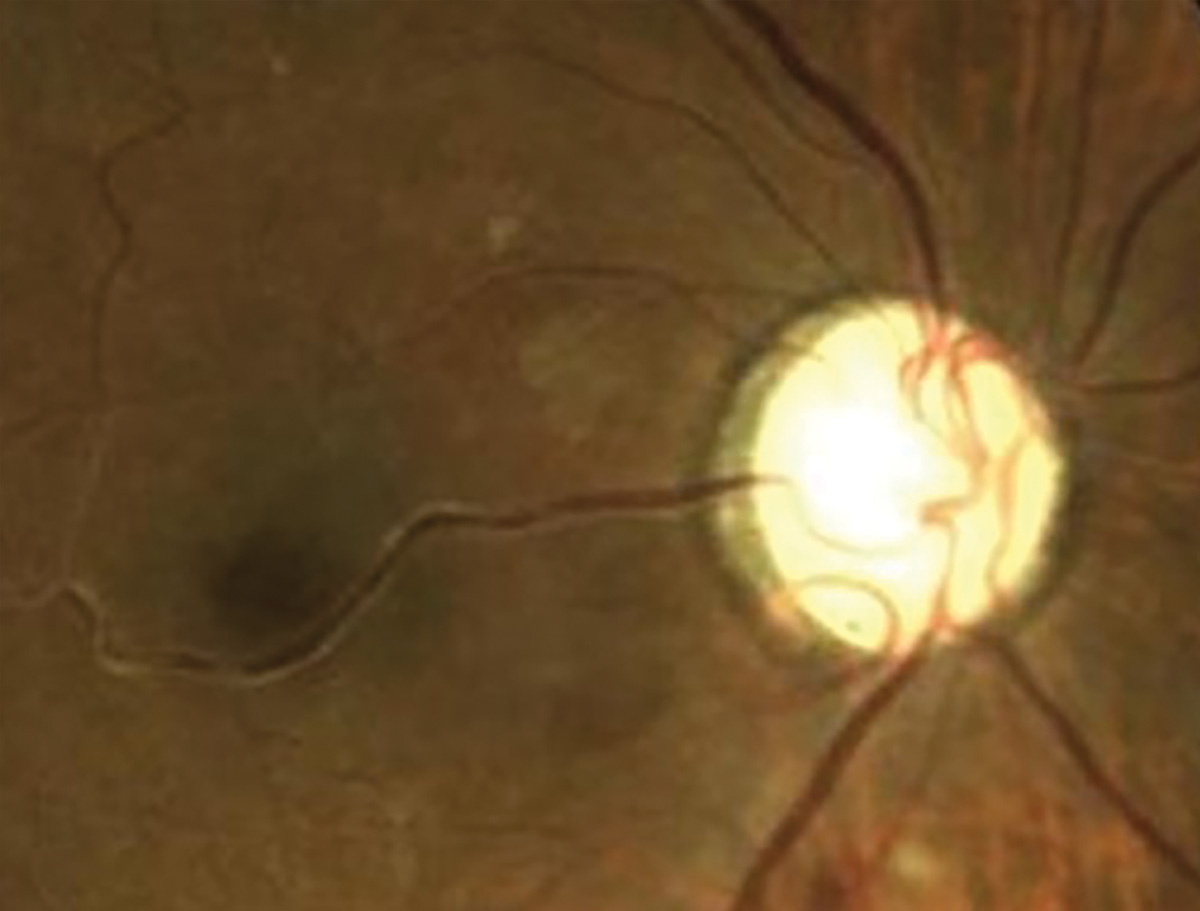
Morning glory disc
 |
| Photo: Mohammad Rafieetary, OD. Click image to enlarge. |
Morning glory disc, a malformation in the spectrum of optic nerve coloboma, with straight vessels radiating from the disc.
Suggested reading:
