To help, Review of Optometry created the following new resource—a photo atlas of ocular diseases broad enough in scope to include conditions both commonplace and rare, benign and worrisome, chronic and acute, across the entire spectrum of eye care.
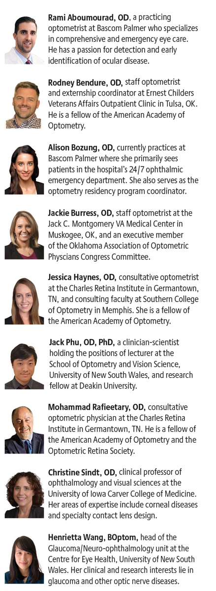
The nine optometrists featured here were instrumental in developing this atlas with us. They shared their time, expertise, patient records and, most of all, their photo libraries to create it. It’s our hope that this issue can give you a quick refresher on the clinical features of many ocular diseases and will become a handy reference guide to aid future patient care responsibilities.
Of course, keep in mind that nearly all conditions can present in a wide variety of ways and what’s depicted here is often just one instance of many. In other words, these photos are representative, but rarely the final word.
This online version of The Photo Atlas of Ocular Disease provides you with a deeper understanding of these conditions in a few ways. First, you’ll be able to view the photos larger and in more detail than in print. Second, we’ll add to it over time, so that new conditions and alternative presentations can be included. Lastly, we will share links to articles from Review’s archives that give in-depth clinical guidance on many of the included diseases and disorders. So, if you see an intriguing photo and would like more context, just follow the links for suggested reading.
I would like to thank all the contributors who made this atlas possible. Their experience and expertise shine through in every photo.
—Jack Persico, Editor-in-Chief
Check out the conditions featured in their respective categories:
- Eyelid Issues: Lumps, Bumps and Beyond
- The Many Mishaps of the Anterior Segment
- Navigate the Retinal Landscape With Confidence
- Assessing the Optic Nerve
- The Physical Manifestations of Glaucoma and What They Signify (CE)
Scroll past the bios to find an index of conditions included.
Our Contributors
Additional photos: Aaron Bronner, OD, Greg Caldwell, OD, Paul Karpecki, OD, Stephanie Fromstein, OD, Mitch Ibach, OD, Nate Lighthizer, OD, Irving Martinez-Navé, OD, Suzanne Sherman, OD |
Use the index below to jump directly to each condition.
Eyelids:
Basal cell carcinoma
Anterior segment:
Epithelial basement membrane dystrophy
Central corneal dystrophy of François
Fuchs’ endothelial corneal dystrophy
Terrien’s marginal degeneration
Peripheral ulcerative keratitis
Conjunctival intraepithelial neoplasm
Giant papillary conjunctivitis
Ocular surface squamous neoplasia
Retina:
Central serous chorioretinopathy
Rhegmatogenous retinal detachment
Retinal angiomatous proliferation
Central retinal vein occlusion
Central retinal artery occlusion
Branch retinal artery occlusion
Intraretinal microvascular abnormalities
Nonproliferative diabetic retinopathy
Proliferative diabetic retinopathy
Optic nerve:
Idiopathic intracranial hypertension
Leber's hereditary optic neuropathy
Anterior ischemic optic neuropathy
