 A 67-year-old white male presented as a new patient for a comprehensive examination in fall of 2008. He complained of decreased distance vision and floaters O.U., but no photopsia.
His last visit to an eye care provider was approximately six years earlier. At that visit, he was informed that his eyes were healthy, and he received an updated eyeglass prescription and glasses.
His current medications include 81mg aspirin q.d.; lisinopril q.d. and atenolol q.d. for hypertension and an unspecified arrhythmia; and vitamins. He reported no known allergies to medications.
Diagnostic Data
Entering visual acuity was 20/50 O.D. and 20/40- O.S. through a refraction of +2.75D0.75D x 095 O.D. and +2.50D0.25D x 075 O.S. Best-corrected visual acuity was 20/25 O.D. and 20/20- O.S. through a refraction of +1.75D 1.25D x 090 O.D. and +1.75D 1.25D x 080 O.S. His pupils were round and reactive to light, with no afferent pupillary defect.
In ambient room light, the right pupil measured 5.5mm and the left pupil measured 4.5mm. This asymmetry remained stable in both bright and dim illumination. Confrontation fields were restricted superiorly by bilateral dermatochalasis.
Slit lamp examination of his anterior segments appeared normal O.U., with clear adnexae and lids, clear corneas, deep and quiet anterior chambers, grade 2+ angles by Van Hericks method and a quiet conjunctiva O.U. Intraocular pressure measured 32mm Hg O.D. and 29mm Hg O.S. at 10:30 a.m.
Through dilated pupils, his crystalline lenses were characterized by moderate nuclear cataracts consistent with his refractive myopic shift and best-corrected visual acuity. There was an irregular ring of exfoliative material on the anterior surface of both lenses outside the pupillary margin, which was not seen without dilation. Vitreous examination demonstrated bilateral posterior separations.
His optic nerves were of normal size and were characterized by a cup-to-disc ratio of 0.55 x 0.75 O.D. and 0.50 x 0.65 O.S. The neuroretinal rims did not follow the ISNT (inferior-superior-nasal-temporal) rule. His vasculature was consistent with mild hypertensive retinopathy O.U. Both maculae had fine retinal pigment epithelium (RPE) granulation in the central foveal avascular zone, with no drusen. His peripheral retinal evaluation was unremarkable.
Given the presence of bilateral exfoliation in conjunction with elevated IOP and suspect optic nerves, we asked the patient to return in two to four weeks for a complete glaucoma work-up.
When he returned for follow-up in two weeks, his IOP measured 35mm Hg O.D. and 31mm Hg O.S. at 8:55 a.m. Pachymetry readings were 576m O.D. and 581m O.S. White-on-white perimetry demonstrated a generalized depression of the superior visual fields; however, his lids were not taped up during the testing. (The technician coached him to keep his eyes open wide during testing.)
Heidelberg Retina Tomograph-3 (HRT-3) imaging of both optic nerves confirmed vertically elongated cups that measured 1.91mm O.D. and 1.89mm O.S. in diameter. Both the Moorfields Regression Analysis and the Glaucoma Probability Score confirmed aberrant optic nerve parameters.
A slit lamp examination of his anterior segment revealed fine exfoliative material at the pupillary margins O.U.
Gonioscopic examination of the anterior chamber angles demonstrated grade 2 to 3 open angles O.U., with a moderate amount of trabecular pigmentation in both eyes. There was a sparse amount of exfoliative material noted in each chamber angle.
Following the evaluation, we made a diagnosis of exfoliative glaucoma O.U. Now, given these findings, how should we proceed with this patient?
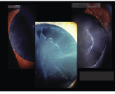
Anterior segment view of exfoliation.
Discussion
In this case, the diagnosis of exfoliative glaucoma was rather straightforward. His IOP was elevated in the absence of excessively thick corneas, exfoliative material was clearly present on the lens and in the angle, and optic nerves were of normal size and show early-to-moderate glaucomatous damage. The visual fields were equivocal, given his lid status. As such, we need to decrease his IOP to a level that hopefully will prevent further optic nerve damage and field deterioration.
To treat this patient properly, we must answer several questions: How do we best achieve IOP reduction? How does the exfoliation factor in? What other factors may be involved? And, what is his prognosis?
First, lets review exfoliation syndrome. General exfoliation syndrome encompasses three clinical conditions: the presence of exfoliative material in the anterior chamber without elevated IOP, exfoliative material with ocular hypertension, and exfoliation and frank glaucoma. Biochemically, the exfoliative material is a byproduct of aberrant extracellular matrix protein production that accumulates in the anterior segment.1 This material may be found in other ocular tissues as well as in the heart, lungs, liver and kidneys.2 Both the exfoliative material and trabecular pigment tend to obstruct the outflow of aqueous humor, which results in elevated IOP. In this respect, exfoliative glaucoma can be considered a secondary glaucoma.
Initial research suggested that exfoliation syndrome was most common in individuals of Scandinavian descent. But, it has since been found in people all over the world.1 Often, exfoliation syndrome initially presents unilaterally in one-third to one-half of casesbut, almost 50% of cases become bilateral within 10 years of onset.1 Approximately 7% of patients with exfoliation syndrome are initially diagnosed with glaucoma, whereas 15% or more may simply have ocular hypertension.3 The likelihood of converting to glaucoma increases with time.
In general, exfoliative glaucoma is usually characterized by higher IOP levels than other forms of open angle glaucoma at the time of diagnosis, and as a result, often is characterized by more advanced glaucomatous optic neuropathy.1,3 Thus, IOP reduction in patients with exfoliative glaucoma must be more aggressive to achieve target IOP, especially if the glaucomatous optic neuropathy is advanced.
Unfortunately, it is often difficult to achieve target IOP in patients with exfoliative glaucoma. Additionally, given the obstructive nature of the underlying mechanical process, exfoliative glaucoma patients typically have greater variations in diurnal IOP curves. Not surprisingly, patients with exfoliative glaucoma are at greater risk of progression than are those who have other forms of open-angle glaucoma, as evidenced in the Early Manifest Glaucoma Study.4
Treatment for exfoliative glaucoma can consist of medications, laser or surgery. Though exfoliation is clearly a risk factor for the development of glaucoma, it actually has protective abilities in the context of management with argon laser trabeculoplasty (ALT).5 While the initial IOP-lowering response from ALT is greater than the response from medical therapy in patients with exfoliative glaucoma, the longevity of this benefit is quite varied and may actually be shorter than in other forms of open-angle glaucoma.3 Surgical trabeculectomy is beneficial for patients with exfoliative glaucoma, but the post-surgical course is often more complex due to zonular weakness (which often accompanies exfoliation syndrome) and a higher incidence of post-operative cataracts.1,3
On one hand, you can approach patients with exfoliative glaucoma as you would other open-angle glaucoma patients. But, on the other hand, these patients are often more difficult to manage and more likely to require additional therapies. Typically, you will have to adjust and modify their management courses more frequently than other open-angle glaucoma patients. Constant vigilance is required when monitoring exfoliative glaucoma patients because long-term IOP control is more difficult to attain.
In this patients case, we initially set a target pressure of 15mm Hg to 16mm Hg O.U. This will require approximately a 50%, or greater, reduction in IOP. While this target may be achieved initially with a prostaglandin, he will likely require ALT in the near future.
We medicated the patient with 1 drop h.s. Travatan Z (travoprost, Alcon) O.U. On initial follow up one month later, IOP readings were 17mm Hg O.D. and 16mm Hg O.S. Because these measurements were very close to target IOP, we elected to keep the patient on monotherapy for the time being. But, we will continue monitoring him for instabilitywhich would not be surprising in this case.
1. Ritch R, Schltzer-Schrehardt U. Exfoliation syndrome. Surv Ophthalmol 2001 Jan-Feb;45(4):265-315.
2. Streeten BW, Li ZY, Wallace RN, et al. Pseudoexfoliative fibrillopathy in visceral organs of a patient with pseudoexfoliation syndrome. Arch Ophthalmol 1992 Dec;110(12):1757-62.
3. Roy FH, Fraunfelder FW. Current Ocular Therapy, 6th ed.
4. Leske MC, Heijl A. Predictors of long-term progression in the Early Manifest Glaucoma Trial. Ophthalmology 2007 Nov: 114(11):1965-72
5. Boland MV, Quigley HA. Risk factors and open-angle glaucoma: classification and application. J Glaucoma 2007 Jun-Jul;16(4):406-18.
