Before the Ocular Hypertension Treatment Study, we measured intraocular pressure without thinking about how central corneal thickness (CCT) affects the accuracy of the IOP measurement. Today, however, we widely accept CCT as an important clinical measurement and take into account its effect on accurate measurement of IOP.
This is especially important when you consider that thin CCT is more likely to be found in patients who have advanced glaucoma, blacks and patients who have normal-tension glaucoma.1 Studies suggest that thinner CCT leads to underestimation of IOP by Goldmann applanation tonometry, resulting in delayed diagnosis and treatment.1,2
The good news: Technology continues to provide new ways to obtain information about the cornea when examining glaucoma patients or suspects and identifying refractive surgery candidates.
Correction Factor
Investigators still debate the IOP correction factor (or correction algorithm) associated with measuring CCT.3 Even with this absence of a generally accepted algorithm for correcting of IOP, as measured by applanation tonometry, clinicians canand shouldwidely apply pachymetry to obtain additional information on an individuals risk of glaucoma, researchers say.3
Thinner CCT has been associated with the degree of glaucoma damage, as indicated by cup-to-disc ratio.4
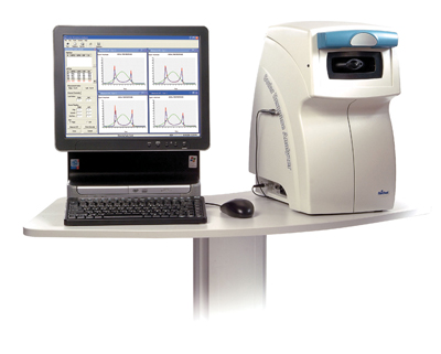 |
| The Ocular Response Analyzer measures corneal hysteresis, which indicates the corneas biomechanical properties. |
New Measurement
Corneal hysteresis (CH) represents a new way of measuring the biomechanical strength, or overall resistance of the cornea. More specifically, it is a measure of viscous damping in the cornea (i.e., absorption of energy from an air pulse). The Ocular Response Analyzer (Reichert) measures both IOP and CH by using a dynamic bi-directional applanation process.5
CH may offer a way to accurately measure the patients true IOP without having to rely on a correction algorithm.6 Also, axial length and CH measurements have been associated with progressive field worsening.4
In one study, researchers compared diurnal variation in CH and IOP and measured CCT with an ultrasound pachymeter.7 IOP, they found, goes through a diurnal fluctuation; it may be highest in the morning and lowest in the afternoon.7 By contrast, CH and CCT measurements tend to remain consistent throughout the day. If the CH measurement changes diurnally, it presumably is due to changes in corneal hydration.8 IOP tends to vary independently of CH or CCT.7
Beyond Glaucoma
The potential applications of CH go beyond glaucoma diagnosis. For example, patients who have keratoconus or Fuchs endothelial dystrophy demonstrate lower CH readings, as do post-LASIK patients.8
CH may join the arsenal of available technology that can help us predict who is most likely to develop corneal ectasia after refractive surgery. Ectasia can occur after otherwise uncomplicated LASIK procedures, even in the absence of apparent preoperative risk factors.9
We already know that corneal topography is a sensitive method for detecting keratoconus earlier.10 This, in turn, helps eye doctors take appropriate measures to improve patients vision and prevent these patients from undergoing refractive surgery.
The ability to assess a patients biomechanical corneal rigidity prior to refractive surgery may also help identify appropriate candidates and predict the outcomes.8 As more patients pursue refractive surgery, time will tell if CH technology can help protect our patients from surgically induced corneal ectasia.
New technologies continue to emerge and join the existing arsenal of technology to allow us to provide better patient care. This includes measuring central corneal thickness, identifying glaucoma suspects and identifying appropriate refractive surgery patients.
Dr. Karpecki is clinical and education conference advisor for Review of Optometry and a contributing editor. Dr. Goodman is clinical director of Advanced Laser Center in Oklahoma City. Neither has any financial interest in the technologies mentioned.
1. Kniestedt C, Lin S, Choe J, et al. Correlation between intraocular pressure, central corneal thickness, stage of glaucoma, and demographic patient data: prospective analysis of biophysical parameters in tertiary glaucoma practice populations. J Glaucoma 2006 Apr;15(2):91-7.
2. Papadia M, Sofianos C, Iester M, et al. Corneal thickness and visual field damage in glaucoma patients. Eye 2006 Apr 28; [Epub ahead of print].
3. Hagerb A, Dave H, Wiegand W. Corneal pachymetry and intraocular pressure. Klin Monatsbl Augenheilkd 2005 Jul;222(7):558-67.
4. Congdon NG, Broman AT, Bandeen-Roche K, Grover D, Quigley HA. Central corneal thickness and corneal hysteresis associated with glaucoma damage. Am J Ophthalmol 2006 May;141(5):868-75. Upub 2006 Mar 9.
5. Reichert Ophthalmic Instruments. Ocular Response AnalyzerUsers Guide.
6. Herndon LW. Measuring intraocular pressure-adjustments for corneal thickness and new technologies. Curr Opin Ophthalmol 2006 Apr;17(2):115-9.
7. Laiquzzaman M, Bhojwani R, Cunliffe I, Shah S. Diurnal variation of ocular hysteresis in normal subjects: relevance in clinical context. Clin Experiment Ophthalmol 2006 Mar; 34(2):114-8.
8. Luce DA. Determining in vivo biomechanical properties of the cornea with an ocular response analyzer. J Cataract Refract Surg 2005 Jan;31(1):156-62.
9. Klein SR, Epstein RJ, Randleman JB, Stulting RD. Corneal ectasia after laser in situ keratomileusis in patients without apparent preoperative risk factors. Cornea 2006 May;25(4):388-403.
10. Jiang HJ, Xie PY. The analysis of corneal topography for keratoconus. Zhonghua Yan Ke Za Zhi 2006 Mar;42(3):231-5.
