 |
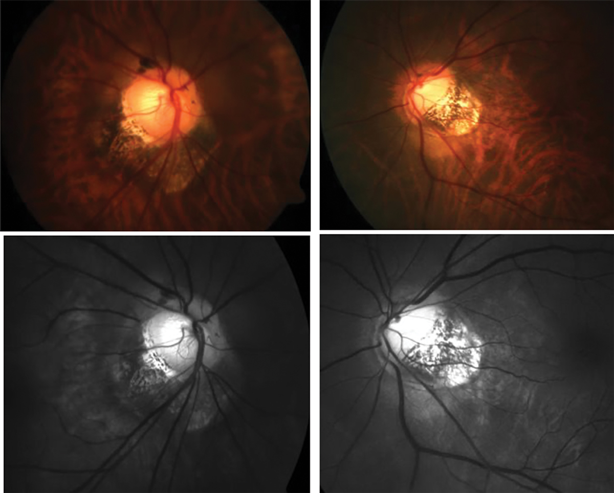
| Traditional fundus photos (top) and red-free images (bottom) show anomalous optic nerve insertion, tilting and torsion and posterior staphyloma in a highly myopic patient. Photo: Andrew Rouse, OD. Click image to enlarge. |
Since some highly myopic eyes can show central and peripheral visual field defects and an abnormally low central visual acuity, researchers thought it may be helpful to better understand optic nerve damage in these patients. In a new report from the Ural Eye and Medical Study conducted in Russia, experts assessed the prevalence of non-glaucomatous optic nerve atrophy (NGOA). High axial myopia showed a relatively high prevalence of NGOA, increasing with longer axial length and wider temporal parapapillary gamma zone.
A total of 5,899 ethnically mixed subjects living near the Ural Mountains, all over the age of 40, participated. NGOA, graded into five arbitrary stages, was characterized by decreased visibility of the retinal nerve fiber layer (RNFL) on fundus photographs and on red-free fundus photographs, neuroretinal rim pallor, abnormally thin retinal arteriole diameter and abnormally thin peripapillary RNFL as measured by OCT.
Findings from this study agree with a previous report on a thinning of the RNFL with wider gamma zone, longer disc-fovea distance and higher systolic blood pressure. The two major parameters with the strongest statistical association with a higher NGOA prevalence were longer axial length and a wider temporal parapapillary gamma zone. For each millimeter of axial elongation and widening of gamma zone, the odds for NGOA increased 7.45-fold and 6.98-fold, respectively.
“Although the biomechanical properties of the retinal nerve fibers have not been explored yet, one may assume that the fibers are not fully elastic or extendable, so that their lengthening may lead to strain and secondary damage,” the authors explained in the study, published in Ophthalmology.
As a corollary, the length of Bruch’s membrane, measured from the foveal center to the end of Bruch’s membrane at the border of gamma zone, does not increase with longer axial length. It indicates that the temporal gamma zone increases the distance between the macular ganglion cells and the optic disc border, so that their axons—the retinal nerve fibers—are elongated by the amount of temporal gamma zone width, the authors pointed out in their article.
“The retinal nerve fibers running in the papillomacular bundle in a primarily straight course to the optic disc can, however, not compensate for the increased distance to the optic disc and may get stretched and potentially damaged in eyes with a large parapapillary gamma zone,” the authors explained.
The associations between a higher NGOA prevalence and higher systolic blood pressure and higher prevalence of diabetes suggest a systemic component in the etiology of NGOA in the highly myopic eyes, either since these two systemic vascular risk factors led to an optic nerve damage independently of axial high myopia, or since diabetes and elevated systolic blood pressure were co-factors in the pathogenesis of a specific high myopia-related damage to the optic nerve fibers,” the authors explained.
Bikbov MM, Iakupova EM, Gilmanshin TR, et al. Prevalence and associations of non-glaucomatous optic nerve atrophy in high myopia. The Ural Eye and Medical Study. Ophthalmology. July 11, 2023. [Epub ahead of print.] |
