 History
A 63-year-old Latino female with a history of Vogt-Koyanagi-Harada (VKH) syndrome and chronic uveitis presented with the chief complaint of a painful right eye.
Her ocular history included various bouts of ocular inflammation, surgical aphakia O.U. and a retinal detachment O.S.
The patient was concerned that her ocular inflammation had started to accelerate because she ran out of topical anti-inflammatory medication several weeks earlier and did not have the financial resources to refill the prescription written by her previous eye care provider. The patients systemic history was remarkable for hypertension. She had no known drug allergies.
Diagnostic Data
Her best-corrected visual acuity was 20/40 O.D. and hand motion O.S. at distance and near. There was a relative afferent defect in her left eye, consistent with previous data that correlated to her complete retinal detachment O.S.
The anterior segment examination uncovered grade 1 cells and flare O.D. We performed a dilated fundus examination of the right eye, but there was no suitable view of the left eye.
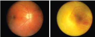
A 63-year-old female with VKH and surgical aphakia O.U. (O.D. left, O.S. right)
Your Diagnosis
How would you approach this case? Does this patient require any additional tests? What is your diagnosis? How would you manage this patient? Whats the likely prognosis?
The Diagnosis
The diagnosis in this case is vitreous prolapse with a recurrence of Vogt-Koyanagi-Harada iritis. VKH disease primarily affects persons who are of Asian, Latino, Native American or Asian Indian descent.1,2 Women appear to be affected more often than men, and the disease may occur at any age.1,2 Experimental data continue to support an autoimmune etiology for VKH disease, which is directed most against an antigenic component of the melanocyte (possibly tyrosinase).1
The clinical diagnosis of VKH disease continues to be based upon physical findings.1 The prognosis of the malady largely depends on what complications develop.1,2
Posterior capsule rupture is the most common serious intraoperative complication of cataract surgery.3-5 A posterior capsular rent (partial tear) is more likely to occur in eyes with small pupils, hard nuclei or pseudoexfoliation syndrome.3-5
Capsular breaks prior to nucleus extraction are dangerous. The surgeon must work to prevent the nucleus from being dislodged into the vitreous cavity. In most circumstances, switching to a manual extraction by sufficiently enlarging the wound to facilitate easy extraction of the nucleus on a lens loop preserves the capsules integrity.3,4
When a capsular rupture occurs during aspiration of the cortex, the status of the vitreous must be observed. If no vitreous is present in the anterior segment, a viscoelastic agent can be injected through the capsular opening to push the vitreous posteriorly. It can be hazardous to remove cortex in the region of a tear, which explains why cortex is sometimes left in eyes with capsular tears.3
If vitreous is present in the anterior segment, a partial vitrectomy should be performed first. If vitreous contacts the corneal endothelium, it will cause endothelial cell death with rapid and permanent corneal decompensation. The misplaced tissue may also obstruct aqueous outflow and produce inflammation with all of the complications from the subsequent exposure.3
Over-inflation of the capsular bag with viscoelastic can also predispose the eye to posterior capsular rupture, with loss of the nucleus into the vitreous.3,4 This is more likely to occur in eyes with long axial lengths or fragile posterior capsules, such as those found in patients who have posterior polar cataracts.3
A ruptured posterior capsule with resultant vitreous prolapse and pupil block angle closure is plausible and has been reported in the literature.5
We treated the patients Vogt-Koyanagi-Harada-based inflammation with atropine 1 % b.i.d. and prednisolone acetate 1 % q.i.d. O.D. for 14 days, and then tapered appropriately.1,2
1. Read RW, Rao NA, Cunningham ET. Vogt-Koyanagi-Harada disease. Curr Opin Ophthalmol 2000 Dec;11(6):437-42.
2. Sheu SJ, Kou HK, Chen JF. Prognostic factors for Vogt-Koyanagi-Harada disease. J Chin Med Assoc. 2003 Mar;66(3):148-54.
3. Kohnen T,
4. Roper-Hall MJ. The lens: nuclear expression of intracapsular cataract surgery. In: Yanoff M, Duker J. Ophthalmology.
