 |
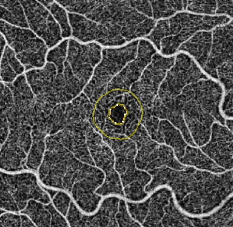
| Beware the possibility of dilating drops affecting accurate measurement of the foveal avascular zone. Photo: Julie Rodman, OD. Click image to enlarge. |
Pupillary dilation is important in conducting an ocular examination or performing intraocular surgery, but a recent study pointed out that mydriatic agents may impact glaucoma diagnostics by altering vascular density measurements.
The study included 20 eyes with primary open-angle glaucoma and 20 control eyes. Eyes underwent fundus imaging before and after instillation of topical 0.5% tropicamide and 2.5% phenylephrine, two commonly used topical mydriasis agents.
The researchers reported a statistically significant decrease in the foveal avascular zone area (from mean 0.29mm2 to 0.25mm2) and the foveal avascular zone perimeter (from mean 2.27mm to 2.09mm), as seen on OCT-A after instillation. They noted that pre- and post-dilation optic nerve head perfusion and flux index were significantly lower in the glaucoma group than the controls.
“It’s long been proposed that glaucomatous eyes have a significant vascular dysregulation and lower ocular perfusion than in normal subjects,” the study authors wrote in their paper. “Pupillary dilation with 0.5% tropicamide and 2.5% phenylephrine resulted in a statistically significant decrease in foveal avascular zone metrics in glaucoma eyes. This observation emphasizes the critical role of pupillary status in interpreting glaucomatous vascular alterations detected by OCT-A.”
Ozturker Z, Kurt RA. Effect of mydriatic administration on retinal hemodynamics in glaucoma: an optical coherence tomography angiography study. J Glaucoma. April14, 2022. [Epub ahead of print]. |
