Age-related macular degeneration (AMD), particularly in the exudative form, is characterized by numerous changes to retinal vessel integrity, but these are typically observed in the aggregate, e.g., overall degenerative changes to the choriocapillaris or loss of vessel density within entire regions of the retinal vasculature. But imaging technology is getting good enough to show changes at the level of individual vessels and that might yield new methods of detecting and monitoring the condition.
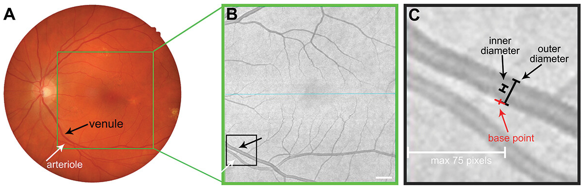 |
| These images from the study show how (A) fundus photography and (B) corresponding en face OCT images were used to identify the largest arteriole and venule closest to the optic nerve head. (C) The outer and inner diameters of each vessel were then measured. Photo: Nivison-Smith L, et al. Clin Exp Optom. Feb 27, 2024. Click image to enlarge. |
The aim of one new study was to use OCT angiography to better understand how the retinal vasculature is altered in eyes with moderate age-related macular degeneration (AMD), as “validation of clinically accessible methods of measuring retinal vascular integrity could provide a more holistic understanding of AMD-related changes to facilitate appropriate care,” for instance with OCT, they wrote.
Included in the study were 46 single eyes of participants with intermediate AMD and 43 single eyes of normal control maculae; all participants were ≥50 years old without diabetes, hypertension or any other systemic vascular disease. Retinal en face OCT images were examined for all of these for arterioles and venules. Arterioles were identifiable in all cases, while venules were identified in 40 AMD patients and 39 controls.
The researchers found good-to-excellent intra- and inter-grader agreement for all en face OCT arteriole and venule diameter measurements. AMD eyes displayed greater arteriolar outer and inner diameters when compared with normal eyes. As well, venular inner diameter was also greater in AMD eyes; outer diameter remained unchanged, suggesting venule remodeling takes place in AMD.
Upon further discussion, the authors elaborate that the greater inner and outer arteriole diameters seen in AMD patients is suggestive of arteriole dilation in early stages of the disease. Other vessel analysis studies corroborate these findings, they explained, showing abnormal retinal arteriole dilation existing in eyes with drusen and reticular pseudodrusen as well as AMD eyes with specific ‘age-related maculopathy susceptibility 2’ genotype. Essentially, these studies suggest that differences in arteriole vessel structure in AMD may be associated with endothelial dysfunction within the vessel wall. Other studies show late AMD patients have circulating endothelial cells and endothelial dysfunction markers via increases in serum, thought to contribute to choroidal neovascularization.
Pertaining to venular remodeling, the authors speculate that “this inner diameter change may reflect outward remodeling of the venule lumen, which has been observed in other systemic vascular diseases, especially considering that AMD and cardiovascular disease share a number of risk factors and potentially a common pathogenesis.” Another alternative could be that the greater venule inner diameter indicates change in the vessel axial light reflex.
This reflex is believed to be a function of vessel diameter and flow velocity of erythrocytes. In this way, disease-induced changes like vessel wall sclerosis or pathology slowing flow velocity leading to more reflexivity and thus a broader axial light reflex. Since venule flow velocity is reportedly unaltered in AMD, venule wall composition alterations could be responsible for altered venule inner diameters in AMD eyes seen here.
As the authors reiterate, “Overall, these results indicate that significant differences in vessel structure exist in intermediate AMD, reinforcing pathophysiological understanding of vascular changes in the early stages of AMD.”
Nivison-Smith L, Faiza A, Roy T, Trinh M. Retinal vessel diameters in intermediate age-related macular degeneration using en face optical coherence tomography. Clin Exp Optom. February 27, 2024. [Epub ahead of print]. |
