 |
| Out of 280 individuals in the study, 52.6% were in the small or large for gestational age groups, while 48.3% were placed in the normal group. Photo: Fieß A, et al. Am J Ophthalmol. Feb. 7, 2024. Click image to enlarge. |
A study published in American Journal of Ophthalmology by German physicians attempted to discover any associations between fetal growth restriction (as well as excessive fetal growth) along with perinatal factors and the optic nerve head development process as manifested in adulthood.
In this retrospective cohort study, the researchers examined individuals born full term between the years 1969 and 2002 at a single institution (University Medical Center Mainz, just outside of Frankfurt). The team had the participants undergo non-mydriatic fundus photography in order to capture images of their optic discs. This was followed by manual measurements. Researchers then examined and analyzed the vertical cup-to-disc (c/d) ratio and optic disc area in relation to each participants’ birth weight relative to their gestational age (SGA: small for gestational age, LGA: large for gestational age), assigning each to one of the following five categories:
• severe SGA: birth weight <3rd percentile
• moderate SGA: 3rd to 9th percentile
• normal SGA: 10th to 90th percentile
• moderate LGA: 91st to 97th percentile
• severe LGA: >97th percentile
The experiment observed 535 eyes from 280 individuals. The mean age for the experiment was 29.74 ±9.23 years (range: 18 to 52), and 144 women and 136 men who were born full term participated in the study. According to the researchers’ multivariate analysis, they found a significant association between a larger vertical c/d ratio and severe SGA status. According to the researchers’ univariate model, placental insufficiency—but no other perinatal factor—was associated with greater vertical c/d ratio.
Interestingly, the researchers did find an indication that a smaller optic disc area was associated with individuals from the moderate SGA group. “No studies have established a direct correlation between lower birth weight for gestational age and optic disc area,” mentioned the researchers in their article on the study. “To date, it has been mainly demonstrated that prematurity is associated with a smaller optic disc.”
The researchers noted that this study was a conducted at a single center and, due to inaccessible contact information from some of the cohorts, the sample was not entirely representative. Additionally, most study participants were of white ethnicity, so the results and conclusions made could only represent this specific population. Also, since this was an exploratory study and researchers did not adjust for multiple testing, they mentioned that more studies would be needed to further understand this area of research.
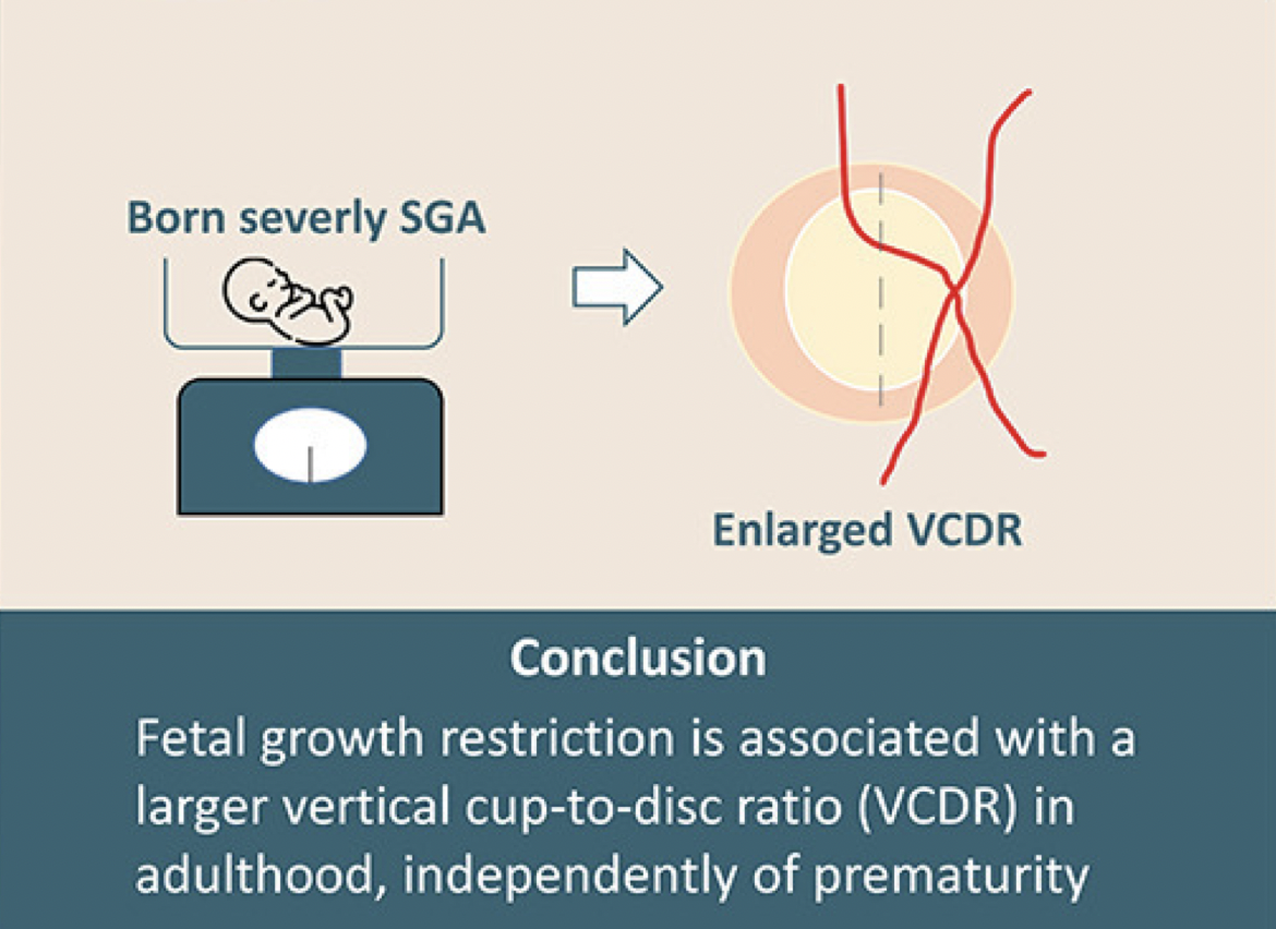
“Our findings provide novel insights into the impacts of fetal growth restriction on the optic nerve head morphology in full-term born individuals, revealing that the vertical cup-to-disc ratio increases in individuals born severely SGA at-term,” concluded the researchers in their paper. “These findings indicate that fetal growth restriction has a lasting impact on the optic nerve head morphology until adulthood and may indicate a lower neuronal reserve for degenerative optic disc diseases.”
Fieß A, Gißler S, Mildenberger E, et al. Fetal growth restriction leads to an enlarged cup-to-disc ratio in adults born at full term. Am J Ophthalmol. Feb. 7, 2024. [Epub ahead of print]. |
