 A 55-year-old Hispanic female presented with blurred vision in her right eye that has persisted for the previous one-and-a- half years. She reported a gradual, painless loss of vision O.D. and no problems O.S.
Her ocular history is unremarkable. Her her medical history is significant for systemic hypertension, for which she takes Diovan (valsartan, Novartis), and asthma, which is controlled with albuterol and Singulair (montelukast sodium, Merck).
Her best-corrected visual acuity was 20/200 O.D. and 20/40 O.S. Confrontation visual fields were full to careful finger counting O.U. Her ocular motility testing was normal. Her pupils were equally round and reactive, with no afferent pupillary defect. Her anterior segment examination was unremarkable.
On dilated fundus exam, her optic nerves appeared healthy with small cups and good rim coloration and perfusion O.U.
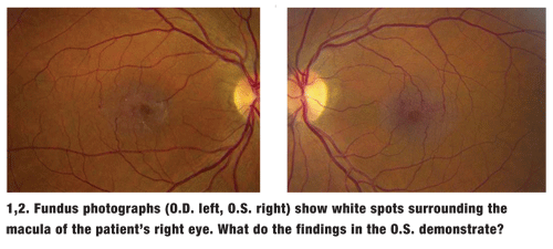
The pertinent fundus changes found in each maculae are illustrated above (figures 1 and 2).
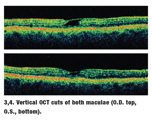
Also, an optical coherence tomography (OCT) scan of both eyes (figures 3 and 4) was performed.
Take the Retina Quiz
1. What do the tiny white spots surrounding the macula in the right eye represent?
a. Fine drusen.
b. Exudate.
c. Cotton-wool spots.
d. Intraretinal crystals.
2. What does the OCT show?
a. Neurosensory retinal detachment.
b. Cystoid space with a retinal drape.
c. Retinal angiomatous proliferations (RAP).
d. Epiretinal membrane with a pseudohole.
3. What is the correct diagnosis?
a. RAP.
b. Cystoid macular edema (CME).
c. Mild hypertensive retinopathy.
d. Idiopathic juxtafoveal retinal telangiectasis (JRT).
4. How should this patient be managed?
a. Observation.
b. Laser photocoagulation.
c. Intravitreal Avastin (bevacizumab, Genentech).
d. Photodynamic therapy (PDT).
For answers, see below.
Discussion
A clinical examination of the eye revealed the presence of fine intraretinal crystals surrounding the right macula. Interestingly, the crystals are not visible in the left eye. In addition, it appears that our patient has a pseudohole O.D.; however, this defect may only be an illusion created by the retinal whitening, which is caused by a combination of retinal thickening and an epiretinal membrane.
While the patient does not have fine intraretinal crystals in her left eye, she does have very obvious dilated retinal capillaries that are easily visible on clinical exam, especially temporal to the fovea.
These findings suggest that our patient has bilateral idiopathic juxtafoveal retinal telangiectasis (JRT), a condition of unknown etiology that is characterized by abnormal retinal vessels that are commonly located temporal to the fovea. As is the case with our patient, abnormal retinal vessels may also surround the fovea.
Patients with JRT develop dilation of the retinal capillaries and telangiectasia of the fine retinal vessels with minimal exudation. They may also develop retinal crystals, as seen in this patient. These crystals are caused by minimal leakage from the telangiectatic retinal vessels.1-2
Another interesting feature of this condition is the small retinal veins around the macula. As they bifurcate from the larger veins, they travel toward the macula and appear to simply stop, which gives the appearance of a blunted end. Actually, the veins do not end; they make a right-angle turn, diving toward the retinal pigment epithelium (RPE). This is evident in both eyes of our patient.
Over time, patients with chronic JRT may develop retinal pigment hyperplasia. When very dense, they can appear as a plaque of pigment. The pigment can even obscure or envelop the vein, making it difficult to visualize the retinal crystals and right-angle venules. Patients with JRT can also develop choroidal neovascularization.
Idiopathic juxtafoveal retinal telangiectasis was first characterized by J.D. Gass and R.T. Oyakawa in 1982, and was later updated by Gass and B.A. Blodi in 1992.1-2 The researchers classified the condition into three types:
Type 1. Individuals with type 1 JRT typically have a unilateral presentation, are more often male than female, and experience disease onset around age 35.1,2 Patients with type 1 probably have a mild form of Coats disease.1,2
In this form, the disease is localized to an area involving 1 to 2 disc diameters (DD) temporal to the fovea, with clearly visible retinal telangiectasis and exudation.
Type 2. This is the most common form of the disease, and is what our patient has. It is bilateral, affects males and females equally, and involves approximately 1DD or less of the temporal foveolar area.1,2
On average, presentation occurs at 55 years of age. In some patients, the telangiectasis can involve the entire parafoveolar area. This form of telangiectasis is associated with minimal to no lipid exudation.
Superficial retinal crystals are seen in approximately 50% of patients.1,2 Additionally, pigment migration is common and often obscures the blunted venules.
Plaque formation is characteristic of type 2 JRT, which often confirms the diagnosis, even when telangiectasis cannot be seen clinically.
Type 3. The least common form of retinal telangiectasis, type 3 JRT typically occurs in older females who develop capillary occlusions. The condition progressively worsens and eventually leads to capillary dropout.
Traditionally, fluorescein angiography (FA) is considered the quintessential diagnostic test for JRT, because it clearly shows areas of retinal telangiectasis that may be difficult to observe clinically.
However, OCT is also used to diagnose JRT. Investigators have even begun to use OCT to categorize the type of JRT by severity.3
In our patient, we see a large central cyst in the right eye with a thin layer of retina draping over it. This is the classic appearance of JRT on OCT imaging. A milder form is observable in the left eye.
Visual acuity in JRT patients ranges from 20/15 to hand motions, with an average of 20/40.1,2 Additionally, no treatment, including laser photocoagulation or photodynamic therapy, has been shown to be effective for JRT patients.
Investigators have now begun experimenting with the anti-VEGF drugs Avastin and Lucentis (ranibizumab, Genentech) for the treatment of macular edema associated with JRT.4 The reports involve small case studies, which have found mixed results. However, greater success has been observed in patients who also develop choroidal neovascularization.5-6
Retina Quiz Answers: 1) d; 2) b; 3) d; 4) c.
1.Gass JD, Oyakawa RT. Idiopathic juxtafoveolar retinal telangiectasis. Arch Ophthalmol 1982 May;100(5):769-80.
2.Gass JD, Blodi BA. Idiopathic juxtafoveolar retinal telangiectasis: update classification and follow up study. Ophthalmology 1993 Oct;100(10):1536-45.
3. Cohen SM, Cohen ML, El-Jabali F, Pautler SE. Optical coherence tomography findings in nonproliferative group 2a idiopathic juxtafoveal retinal telangiectasis. Retina 2007 Jan;27(1):59-66.
4. Moon SJ,
5. Ruys J, De Laey JJ, Vanderhaeghen Y, et al. Intravitreal bevacizumab (Avastin) for the treatment of bilateral acquired juxtafoveal retinal telangiectasis associated with choroidal neovascular membrane. Eye 2007 Aug 10; [Epub ahead of print].
6. Jorge R, Costa RA, Calucci D, Scott IU. Intravitreal bevacizumab (Avastin) associated with the regression of subretinal neovascularization in idiopathic juxtafoveolar retinal telangiectasis. Graefes Arch Clin Exp Ophthalmol 2007 Jul;245(7):1045-8.
