 A 39-year-old Hispanic male presented with mild pain and poor vision in his left eye that had persisted for three weeks. The patient also reported blurred vision in his right eye that had persisted for nearly a week.
The patient was HIV-positive and had stopped taking his medications. The patient reported that he had not seen his physician in more than a year, so he was unaware of his CD4 count or viral load.
On examination, his best-corrected visual acuity measured 20/60 O.D. and hand motion (HM) O.S. Confrontation visual fields were full to careful finger counting in his right eye, but severely constricted in his left eye—HM only. His right pupil was round and sluggishly reactive to light, and his left pupil was irregularly shaped with minimal reaction to light. There was a 1-2+ left afferent pupillary defect on reverse testing.
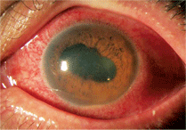
1. Dilation revealed posterior synechia in our patient’s left eye.
The anterior segment exam revealed 1+ cell in the right anterior chamber and 3+ cell and flare in the left anterior chamber. There was also a small hypopyon visible in the left eye in addition to a significant posterior synechia present in the left eye (figure 1). Intraocular pressure measured 14mm Hg O.U.
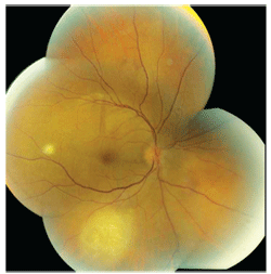
2. This montage fundus photograph of the right eye shows a focal white lesion
temporal to the macula.
Dilated fundus exam was significant for 1+ vitreous cells in the right eye and 3+ cells in the left eye. The right optic nerve had a small cup with good rim coloration and perfusion. However, we could not view the left optic nerve due to the presence of vitritis and posterior synechia.
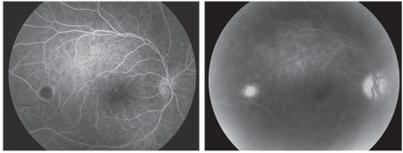
3, 4. Early- (left) and late-stage (right) fluorescein angiogram images show the
posterior pole, macula and peripheral retina of the patient’s right eye.
The right eye showed a diffuse, white plaquoid mass involving the macula and superior arcade. We also noted a small, white focal mass temporal to the macula as well as a larger mass located along the inferior arcade that appeared to be at the level of the retinal pigment epithelium (RPE) and choroid (figure 2). The optical coherence tomography scan of both eyes was normal. We also performed early- and late-stage fluorescein angiogram (FA) of the right eye (figures 3 and 4).
Take the Retina Quiz
1. How would you classify this patient’s inflammation?
a. Active retinitis.
b. Posterior uveitis.
c. Active chorioretinitis.
d. Panuveitis
2. What diagnostic tests are necessary to help establish a diagnosis?
a. Toxoplasma titre.
b. Fluorescent treponemal antibody absorption (FTA-Abs) and rapid plasma reagin (RPR).
c. Cytomegalovirus (CMV) titre.
d. All of the above.
3. Based on the exam findings, what is the most likely diagnosis?
a. CMV retinitis.
b. Herpes zoster retinitis.
c. Active syphilis chorioretinitis.
d. Toxoplasmosis.
4. What is the most appropriate management option?
a. Topical steroids and cycloplegics.
b. Triple sulfa.
c. Intravenous acyclovir.
d. Intravenous penicillin.
5. What additional testing should be recommended?
a. Purified protein derivative (PPD).
b. Visual fields.
c. Ultrasound.
d. Lumbar puncture (LP).
For answers, see below.
Discussion
Our patient had fairly severe uveitis in both eyes. Uveitis is categorized into anterior, posterior and intermediate. However, when the entire uveal track is inflamed, it is called a panuveitis. The presence of cell and flare in the anterior segment and vitreous cells as well as the existence of chorioretinal lesions indicates complete uveal track involvement. When this occurs, the condition is most often systemic in nature.
So, what is causing such a uveitis in our patient? The differential diagnosis can be pretty extensive. Toxoplasmosis is a possibility, but it often presents unilaterally. Tuberculosis is also a possibility, but in this modern day of medicine, it is merely an outside possibility. The presentation also could be caused by sarcoidosis or other complications secondary to HIV. Finally, you cannot overlook the great masquerader––syphilis.
We initiated a medical work-up, which included a PPD, chest X-ray and an MRI. We also conducted a blood work-up, which consisted of an FTA-Abs, RPR and angiotensin-converting enzyme (ACE) tests. We obtained an ultrasound of the left eye to rule out the presence of a mass or retinal detachment. It showed no retinal detachment; however, we noted dense vitreous opacities and marked fundus thickening. We started the patient on Pred Forte (prednisolone acetate, Allergan) q.h. O.U. and scopolamine 0.25% b.i.d. O.U.
The patient returned the following day. He did not undergo the MRI; however, the FTA-Abs came back as being “reactive” and the RPR was positive. These tests indicated that our patient had active syphilis.
Syphilis is caused by Treponema pallidum, a highly infectious spirochete. It has always been, and continues to be, considered a great masquerader in medicine. Following the introduction of penicillin in the early 20th century, the incidence of syphilis declined rapidly. This pattern changed in the late 1980s, however, when a steady resurgence was noted due to changes in sexual practice and the rise of the AIDS epidemic.1 From the late 1980s to the start of the 21st century, safer sexual practices spurred another rapid decline of in syphilis cases. Unfortunately, by 2003, syphilis began making a resurgence, suggesting that more people were engaging in unprotected sex once again.1
Historically, syphilis has been divided into three clinical stages: primary, secondary and tertiary.
• Primary syphilis. This stage is characterized by a chancre on the mucous membranes that usually appears within the first three weeks of inoculation. Typically, ocular involvement is not seen in the primary phase.
• Secondary syphilis. The second stage of syphilis occurs anywhere from two months to three years after inoculation. In this stage, patients may present with anterior uveitis, retinochoroiditis, neuroretinitis and optic neuropathy.2 Nevertheless, it is possible that patients may pass through this stage without experiencing any noticeable symptoms.
• Tertiary syphilis. In this stage, the immune system breaks down and treponemes are detectable in various tissues, including the aorta and brain. Many of the same ocular findings in secondary syphilis are also seen in tertiary syphilis, with the addition of optic atrophy and Argyll Robertson pupil.2
Laboratory tests may be necessary to establish a diagnosis of syphilis. Nonspecific tests, such as Venereal Disease Research Laboratory (VDRL), RPR and radiosensitivity (RST), will be positive in active cases of syphilis and can be used to help determine the effectiveness of systemic treatments. Additionally, specific tests, such as FTA-Abs and microhemagglutination-Treponema pallidum (MHA-TP), are usually positive in patients even after effective treatment. An LP that includes an evaluation of cerebrospinal fluid is indicated for patients suspected of having secondary or tertiary syphilis.
Our patient had extensive syphilitic involvement, as evidenced by the panuveitis. Of particular interest is the faint placoid lesion that extends from the macula to above the superior arcade in his right eye. This lesion is consistent with “syphilitic posterior placoid chorioretinitis,” which was first described by J. Donald M. Gass, M.D., and associates in 1990.3 Gass suggested that such presentations appear as yellowish, ill-defined, placoid lesions that are confluent in the posterior pole or mid-periphery of the fundus. They usually feature a faded center and stippled hyperpigmentation of the RPE and can coalesce to become large, confluent lesions. Syphilitic posterior placoid lesions are most commonly seen in immunocompromised patients. In this case, our patient had a single, large placoid lesion that stained late on FA.
The other lesions seen in our patient were consistent with active chorioretinitis, which can be accompanied by a variable amount of vitreous inflammation. These lesions also may be associated with superficial hemorrhages, retinal vasculitis, and disc edema; however, these features were not present in our patient.
Our patient was admitted to a local hospital to undergo treatment with IV penicillin and an LP to rule out neurosyphilis. Subsequently, we lost him to follow-up. When he returned two and a half years later, his visual acuity measured 20/80 O.D. and 20/200 O.S.
There was no inflammation present in either eye. His right macula appeared completely normal. There were two focal nonspecific chorioretinal scars (a smaller scar temporal to the macula and a larger scar along the inferior arcade, which was the same size as the active lesion seen two and a half years earlier). Posterior synechia was still present in the left eye; however, he did dilate sufficiently for a clear view into the retina. The left optic nerve was pink and healthy, and the macula showed a mild epiretinal membrane. We also noted a large, inactive chorioretinal scar along the superior arcade that extended anteriorly to the peripheral retina.
We asked him to return for visual field testing and a multifocal electroretinogram. He has yet to return to our office.
1. Kerani RP, Handsfield HH, Stenger MS, et al. Rising rates of syphilis in the era of syphilis elimination. Sex Transm Dis. 2007 Mar;34(3):154-61.
2. Knox DL. Retinal Syphilis and Tuberculosis. In: Ryan SJ, Schachat AP, Murphy RP (eds). Retina, vol. 2. Medical Retina, 4th ed. St. Louis: Mosby; 2006:1711-9.
3. Gass JD, Braunstein RA, Chenoweth RG. Acute syphilitic posterior placoid chorioretinitis. Ophthalmology. 1990 Oct;97(10):1288-97.
