 |
|
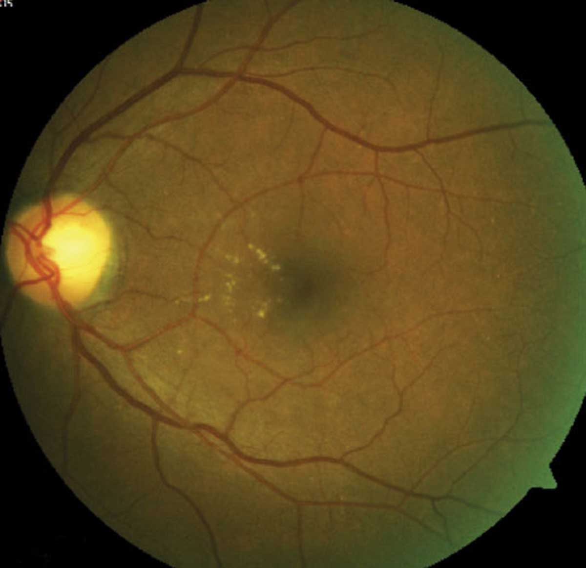
A recent study found that the counts of intercapillary spaces in seven significant sectors were feasible to infer VA reduction in DR. Click image to enlarge. |
Authors of a recent study proposed an intercapillary space spectrum that contains both healthy intercapillary areas and pathophysiological NPAs on OCT-A images, which can be used to evaluate the processes of capillary nonperfusion automatically and objectively. They investigated the clinical significance of intercapillary spaces on swept-source OCT-A images in diabetic retinopathy (DR).
A total of 110 eyes of 110 patients with DR without macular edema were evaluated with swept-source OCT-A images centered on the fovea. Automatic image processing of the superficial slab images allowed the authors to define the areas encircled by retinal vessels as intercapillary spaces within the central 2mm circle. They evaluated how the quantitative parameters of intercapillary spaces are associated with logMAR and feasible to diagnose diabetic macular ischemia.
The study found that numbers of intercapillary spaces in several sectors of the outer rings were significantly associated with VA reduction, which suggests that parafoveal ischemic changes in the superficial vascular layer impact acuity, the authors explained.
“Bipolar cells and subsequent ganglion cells from cone photoreceptors are centrifugally displaced and anatomically correspond with the FAZ and the superficial vascular layer in the parafovea, respectively,” they explained. “Diabetes leads to damages in both capillaries and might contribute to the impairment in ganglion cells and resultant VA decrease, at least in part.”
All significant sectors, in which capillary nonperfusion was highly associated with VA reduction, were located in the superior hemifield rather than in the inferior hemifield. The authors noted that previous studies demonstrated that the retinas in the superior quadrant tend to have greater thicknesses on OCT imaging and higher perfusion metrics in the superficial OCT-A images than those in the inferior quadrant in healthy eyes.
“This finding suggests that retinal parenchyma in the superior areas needs greater amounts of oxygen and glucose, which are supplied by denser capillaries,” the authors concluded. “In addition, the inner retinal layers of the superior areas are farther from the choroidal nourishment. In contrast, there were no differences in the capillary flow density between the superior and inferior quadrant in diabetic eyes. These publications may allow us to speculate that capillary nonperfusion in the superficial layer promotes ganglion cell dysfunction more significantly in the superior hemifield than in the inferior hemifield.”
Terada N, Murakami T, Ishihara K, et al. Clinical relevance of parafoveal intercapillary spaces and foveal avascular zone in diabetic retinopathy without macular edema. Invest Ophthalmol Vis Sci. 2022;63(12):4. |
