 Q: I’ve heard about a recent study that links Alzheimer’s disease and the eye. I have at least a couple of patients who I suspect have Alzheimer’s. Are there ocular findings I can look for in these patients?
A: Most certainly. “Everybody says the eye is a window to the brain. But the eye is not only a window, the eye is actually part of the brain,” says retina specialist Clement Trempe, M.D., professor of optometry at the New England College of Optometry and head scientist with the Center for Healthy Aging at the New England Eye Institute.
More than 5.3 million Americans have Alzheimer’s, and it’s the sixth-leading cause of death, according to the Alzheimer’s Association.
One of the main hurdles in diagnosing Alzheimer’s is that it’s not easy or cheap to diagnose. The only definite way to diagnose the disease is with an autopsy. Short of that, brain scans, such as computed tomography (CT) or magnetic resonance imaging (MRI), can be performed.
But an MRI costs $4,000, Dr. Trempe says. “Little progress will be made in the treatment of Alzheimer’s disease unless we find a readily available, easy, repeatable, non-invasive way to diagnose the disease early,” he says.
To that end, optometrists can perform the comprehensive eye exam that they do every day using existing, readily available equipment to detect the ocular signs of Alzheimer’s early on in the disease process, Dr. Trempe says.
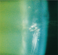
This is the type of cortical cataract that may indicate early Alzheimer’s disease.
“Optometrists are located in every part of the country. They see more than 50 million patients every year,” he says. “They are in an ideal position to detect the early signs of Alzheimer’s disease in the general population.”
These early signs, or biomarkers, can be easily overlooked because the pathological findings associated with Alzheimer’s disease are similar to, and overlap with, the findings associated with common age-related eye diseases.
These biomarkers include cortical cataract, retinal nerve fiber layer thinning and drusen.
• Cortical cataract. “Amyloid beta deposits are seen in the cortex of the lens in patients with Alzheimer’s disease,” Dr. Trempe says. These are the same type of proteins that are a hallmark of Alzheimer’s when found in the brain. The amyloid beta proteins that form plaques in the brain and impair cognitive function also build up in the peripheral cortical area of the crystalline lens, ultimately forming an unusual “supranuclear” cataract that is different from more familiar, age-related cataracts.1
Upon first glance, though, this cataract may look like a garden-variety cortical cataract. The difference is that the amyloid beta deposits form observable amyloid fibrils that ultimately result in cataract. “You have to use a narrow slit lamp beam at high [40x] magnification to spot the fibrils,” Dr. Trempe says, although he acknowledged that even when pointed out, these fibrils can be hard to visualize.
Another clue: The amyloid beta protein accumulations cause swelling in the periphery of the lens early in the disease process, “which in turn produce a specific type of coma aberration,” Dr. Trempe says. “This distortion can be measured highly accurately using the same instruments employed for corneal refractive surgery.”
• Retinal nerve fiber layer thinning. The retina is a direct extension of the brain, Dr. Trempe says. So, “when you see something changing in the eye, it’s also changing in the brain. Glaucoma, like Alzheimer’s, is a neurodegenerative disease, but it manifests on the optic nerve.” Indeed, retinal amyloid beta deposits are strongly implicated in both Alzheimer’s disease and glaucoma.
Various researchers, including Dr. Trempe and colleagues, have found specific deterioration of the retina in patients with Alzheimer’s. In particular, they found significant thinning of the peripapillary retina nerve fiber layer that was most pronounced in the superior quadrant.2 This corresponds to visual field defects predominantly in the inferonasal and inferotemporal arcuate regions in Alzheimer’s patients.3
Using clinical examination and scanning laser ophthalmoscopy, other researchers have found a reduction in the number of optic nerve fibers in patients with Alzheimer’s disease. These patients were also three times more likely to have larger cup-to-disc ratios.4 In many cases of Alzheimer’s disease, the optic nerve showed predominant loss of the largest class of retinal ganglion cells (M-cells) that contribute large caliber fibers to the optic nerve.5
• Drusen. Amyloid beta deposits have also been found in some drusen. Specifically, amyloid beta was found in drusen from some retinas with age-related macular degeneration, but not in drusen from normal retinas, according to researchers from Scheie Eye Institute and the Alzheimer’s Disease Center in Philadelphia.6 Amyloid beta drusen were most numerous in eyes with geographic atrophy at the edges of the atrophy, the region at risk for further degeneration. “The finding of similarities in the pathogenesis of AMD and Alzheimer’s disease suggests that much can be learned about one disease from the other,” the researchers concluded. “Anti-amyloid beta therapies currently under development for Alzheimer’s disease may also prove useful for AMD.”6
Q: Suppose I observe some of these findings; is there anything I can do? Should I discuss it with the patient and their family, or just refer the patient to a neurologist?A: “There’s a lot of things the optometrist can do,” Dr. Trempe says. The key is to act early in the pre-symptomatic stage. By the time Alzheimer’s is manifest, it’s usually too late to do anything about it.
“If we can delay the onset of Alzheimer’s by five years, we can cut in half the number of people who will die with the disease,” he says.
For example, a patient with early cortical cataracts could very likely have concurrent inflammatory systemic disease. Spend 15 minutes with him to ask and discuss lifestyle concerns.
Alzheimer’s is a degenerative disease. But, “we’re all degenerating. You just don’t want to degenerate too fast,” Dr. Trempe says. “If the patient smokes two packs a day, he’ll degenerate faster. If he has a lousy diet, doesn’t eat fish, never eats fruit, is 70 years old, then it’s almost no wonder that he’s losing his memory.”
In addition, cognitive deficits can be detected in patients long before a clinical diagnosis of Alzheimer’s can be reached, Dr. Trempe says. “For example, defects in visual memory can be identified decades before overt symptoms of Alzheimer’s disease appear. These problems of visual memory can be diagnosed using the readily available, low-cost Benton Visual Retention Test.”
Optometrists in many states can order blood tests, Dr. Trempe says. Tests for high levels of homocysteine, C-reactive protein and white blood cell count all may indicate inflammatory-type diseases. For instance, people with elevated levels of homocysteine in the blood had nearly double the risk of developing Alzheimer’s disease, according to the Framingham Study.7
The solution is as simple as eating a healthy diet and taking vitamin supplements, Dr. Trempe says. “Homocysteine can be reduced by taking folic acid, vitamin B6 and vitamin B12.”
Lastly, if your clinical examination and discussion with the patient leads you to refer the patient to a neurologist, be sure to make clear to the neurologist that you’ll continue to follow the patient, Dr. Trempe says. Several medications, such as Aricept (donepezil, Eisai/Pfizer) or Namenda (memantine, Forest Pharmaceuticals), are now available to slow the progression of Alzheimer’s.
Continue to monitor the patient for ocular and cognitive signs and keep the neurologist informed.
1. Goldstein LE, Muffat JA, Cherny RA, et al. Cytosolic beta-amyloid deposition and supranuclear cataracts in lenses from people with Alzheimer’s disease. Lancet. 2003 Apr 12;361(9365):1258-65.
2. Berisha F, Feke GT, Trempe CL, et al. Retinal abnormalities in early Alzheimer’s disease. Invest Ophthalmol Vis Sci. 2007 May;48(5):2285-9.
3. Trick GL, Trick LR, Morris P, Wolf M. Visual field loss in senile dementia of the Alzheimer’s type. Neurology. 1995 Jan;45(1):68-74.
4. Danesh-Meyer HV, Birch H, Ku JY, et al. Reduction of optic nerve fibers in patients with Alzheimer disease identified by laser imaging. Neurology. 2006 Nov 28;67(10):1852-4.
5. Sadun AA, Bassi CJ. Optic nerve damage in Alzheimer’s disease. Ophthalmology. 1990 Jan;97(1):9-17.
6. Dentchev T, Milam AH, Lee VM, et al. Amyloid-beta is found in drusen from some age-related macular degeneration retinas, but not in drusen from normal retinas. Mol Vis. 2003 May 14;9:184-90.
7. Seshadri S, Beiser A, Selhub J, et al. Plasma homocysteine as a risk factor for dementia and Alzheimer’s disease. N Engl J Med. 2002 Feb 14;346(7):476-83.
