Ocular manifestations occur in approximately 15% of patients with systemic lupus erythematosus (SLE). However, children with SLE may have a higher risk of ocular complications.1
SLE is a potentially fatal disease that involves significant ocular findings. This chronic autoimmune inflammatory disease can affect the skin, kidneys, joints, nervous system, blood, eyes and other organs. The disease is considered an imitator disorder because its symptoms and clinical course vary and mimic other conditions. SLE is predominant among females and is characterized by periods of flares and remissions, varied multi-system involvement and autoantibodies that act primarily against nuclear antigens.
The five-year survival rate of patients with SLE has increased from 50% in the 1950s to between 91% and 97% today. Mortality rates are higher among SLE patients who develop infectious complications, seizures, lupus nephritis and renal failure.2
 |
|
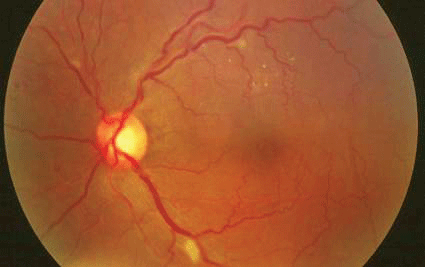
Cotton-wool spots are the most common finding of retinopathy, an ocular manifestation of SLE. |
Etiology
SLE is a complex disease with an unknown etiology. The disease cascade is likely activated by a combination of genetic, environmental and hormonal factors that are initiated by a trigger, such as a bacterial or viral infection. One study found that 11% of patients with SLE had an inherited complement protein defect.5 The concordance rate of SLE is 2% to 9% in dizygotic twins and 24% to 65% in monozygotic twins.6 At least four susceptibility genes and up to 100 genes are estimated to be necessary for development of the disease.6,7 While a female predilection is clear, endogenous sex hormones have an unknown role in disease predisposition and activity.3
Ultraviolet light, infections and medications, including Apresoline (hydralazine, Novartis) and Procan, (procainamide, Erfa Canada), can trigger flares or exacerbations of SLE. Flares are also associated with periods of hormonal changes, such as pregnancy and initiation of hormone replacement therapy.
The prevalence of SLE is approximately 1 in 2,000, and frequency varies by ethnicity.8 An estimated 1.5 million Americans have some form of lupus. Of these, 90% are female, and 80% develop SLE between ages 15 and 45.9 SLE is also two to three times more prevalent in blacks, Asians, Hispanics and Native Americans.9
The pathology of SLE consists of vascular abnormalities and inflammation. These result in immune complex deposition, occlusive vasculopathy and vasculitis.
Patients initially experience a flu-like syndrome, including fatigue, fever, weight loss, and muscle and joint pain. As SLE progresses, complications become more diverse and severe. Among them: a malar rash on the cheeks, rashes elsewhere and alopecia (see Frequency of SLE Involvement Within Systems).
|
Frequency of SLE Involvement Within Systems |
| System | Description | Frequency |
| Hematological | Hemolytic anemia, leukopenia, lymphopenia, thrombocytopenia |
95% |
| Joints | Non-erosive arthritis |
95% |
| Cutaneous | Malar rash, discoid rash | 80% |
| Pulmonary | Pleurisy |
65% |
| Nervous | Generalized seizures, psychosis, organic brain syndrome |
60% |
| Renal |
Related to immune complexes localized in kidneysnephritis, nephrotic syndrome, renal failure |
30% to 55% |
| Gastrointestinal | Nausea, vomiting and anorexia |
20% |
| Ocular | Dry eye, vasculitis, choroidopathy, optic neuropathy |
15% |
|
Incidence of Signs and Symptoms Associated with SLE |
| Symptom | Description | Incidence |
| Arthralgia | Joint discomfort | 95% |
| Arthritis |
Involves two or more swollen joints associated with inflammation (does not affect surrounding bone) |
90% |
| Fever | More than 100 degrees | 90% |
| Fatigue | May be prolonged or extreme in nature | 81% |
| Skin rashes | Raised red patches | 74% |
| Anemia | Hemolytic/leukopenia/lymphopenia | 71% |
| Kidney | Proteinuria or other abnormal elements in urine | 50% |
| Pleurisy | Pain in chest upon breathing; inflammation of the lining of the lung | 45% |
| Malar ("butterfly") rash | Rash pattern noted across cheeks and nose | 42% |
| Photosensitivity | Abnormal sensitivity to the sun or light, resulting in development/increase in skin rash | 30% |
| Alopecia | Hair loss | 27% |
| Abnormal blood clotting problems | Associated with hemolytic anemia, leukopenia and thrombocytopenia | 20% |
| Raynaud"s phenomenon | Fingers turning white and/or blue; associated with cold | 17% |
| Seizures | Convulsive in nature | 15% |
| Ulcers | Mouth and nose; generally painless | 12% |
|
Table information adapted from www.lupus.org/education/sympt.html. |
A diagnosis of SLE is based on four of 11 diagnostic criteria established by the American College of Rheumatology (see Diagnostic Criteria for SLE). Symptoms do not need to be concurrent. Serology may be beneficial but is not necessary for diagnosing SLE.
|
Diagnostic Criteria for SLE |
| Criterion | Description |
| 1. Malar rash | Flat or raised persistent rash across the cheeks and nose |
| 2. Discoid rash | Raised red patches on the skin with associated scaling; atrophic scarring may arise |
| 3. Photosensitivity |
Abnormal sensitivity to light, resulting in development or increase of a skin rash |
| 4. Ulcers |
Noted in mouth or nasal passage |
| 5. Arthritis |
Two or more peripheral joints must be involved; non-erosive to bone but characterized by tenderness and swelling |
| 6. Serositis |
Inflammation of the lining of the lungs or heart |
| 7. Renal disorder |
Protein in urine and/or abnormal elements in the urine |
| 8. Neurologic disorder |
Psychosis and/or seizures in the absence of metabolic conditions or drugs that might be causative |
| 9. Hematologic disorder | Leukopenia or lymphopenia noted on two or more occasions; hemolytic anemia or thrombocytopenia in the absence of causative drugs. |
| 10. Immunologic disorder |
False positive on syphilis test (i.e., venereal disease research laboratory); positive anti-double stranded anti-DNA test, anti-Sm test, lupus anti-coagulant or antiphospholipid antibody |
| 11. Antinuclear antibody (ANA) |
Positive test for ANA in the absence of drugs known to cause a positive response |
|
The original eleven diagnostic criteria for classifying patients with SLE were revised in 1997.17,18 |
Lab tests that may help in the diagnosis and management of SLE include:
Red blood cell count, hemoglobin, hematocrit and iron levels to measure for anemia, which occurs in approximately 50% of patients with SLE during the course of their disease.10
White blood cell count to measure for leukopenia, which occurs in 20% to 30% of SLE patients.8
Platelet count to measure for thrombocytopenia, which occurs in 20% of SLE patients.8
Erythrocyte sedimentation rate (ESR) and C-reactive protein (CRP) to measure inflammation. CRP has a more immediate response and will rise and fall more quickly than ESR with both the increase and the decrease of inflammation.
Antinuclear antibody (ANA) to rule out SLE. This screening test has a high sensitivity and low specificity. Antinuclear antibodies are present in more than 95% of SLE patients at some point in their disease.3
Anti-Sm, anti-nDNA and antiphospholipid antibodies (APLs). These are specialized tests adminstered when ANA is positive.
Urinalysis, red and white blood cell counts, and cellular casts to test for kidney disease. Creatinine concentration measurement and clearances can be used to evaluate kidney function, and kidney biopsy can help physicians determine the extent and type of inflammation.
Urinalysis, complete blood count, complement (C3 and C4) serum levels, and anti-nDNA to measure disease activity. Low complement levels indicate a significant immune reaction usually with kidney involvement.
Treatment
There currently is no cure for SLE. So, the goal of therapy is to alleviate symptoms, prolong remissions, reverse immune dysregulation and prevent organ damage.
Specific therapy depends on the individual and severity of disease. First-line drugs include nonsteroidal anti-inflammatory drugs (NSAIDs) and corticosteroids, which have long been used to control symptoms. Second-line drugs include disease-modifying anti-rheumatic drugs (DMARDs), such as Plaquenil (hy-droxychloroquine, Sanofi); Neoral (cyclosporine, Novartis) and Imuran (azathioprine, GlaxoSmith-Kline), which suppress the immune system; and biologic response modifiers (BRMs), such as Kineret (anakinra, Amgen), which influence the activity of cytokines.
Corticosteroids and hydroxychloroquine may cause secondary ocular complications. Chronic use of oral corticosteroids to treat systemic disease increases patients risk of developing cataracts, most commonly posterior subcapsular (PSC) cataracts. So, we must monitor patients for lenticular changes and educate them about the possible development of cataract and the associated visual consequences.
Elevated IOP more commonly occurs with topical corticosteroid use than with oral corticosteroids, but IOP measurements should still be performed at each visit. If IOP increases significantly, patients may require more frequent IOP measurements along with visual field testing and/or nerve fiber layer analysis.
We also must carefully monitor patients taking hydroxychloroquine or chloroquine for retinopathy, which is partly dose-dependent. Retinopathy may start out as subtle, asymptomatic and reversible mottling of the macular pigment. If left unrecognized, it may progress to a bulls-eye maculopathy with profound, irreversible vision loss.
Visual field testing is very important, as it may detect ocular complications associated with hydroxychloroquine before retinopathy becomes visible. Thus, central visual field testing and a dilated fundus examination should be performed at least once a year, or every six months for patients who have been taking a dose greater than 6.5mg/kg each day for more than five years. Multifocal electroretinography (mfERG) may be used to confirm a retinal etiology when you detect visual field defects. One study demonstrated that mfERG detected retinal physiological changes earlier than visual acuity testing, Ishihara color vision analysis or Amsler grid testing in patients treated for rheumatoid arthritis (RA), SLE or localized atypical scleroderma.11
Ocular Manifestations
The ocular complications of SLE are variable. They appear more frequently in patients who also have concurrent systemic disease.
Keratoconjunctivitis sicca (KCS) is the most common ocular manifestation of SLE. Symptoms of KCS include chronic dryness, intermittently blurred vision and, when severe, ocular discomfort or pain. Ocular findings include conjunctival staining, punctate epithelial keratitis (PEK), diminished tear meniscus and decreased tear break-up time. Staining with rose bengal or lissamine green, Schirmers test and phenol red thread test can be used to assess the presence and severity of KCS.
Autoimmune disorders that may accompany SLE can compromise the lacrimal system. These include Sjgrens syndrome, which is a multi-factorial condition comprised of dry eyes (KCS), dry mouth (xerostoma) and systemic conditions, such as SLE, RA, systemic sclerosis, psoriatic arthritis, Hashimotos thryroiditis and juvenile chronic arthritis.
Generalized orbital inflammation in the form of acute proptosis, lid edema, and/or limited ocular motility may also occur in SLE patients. Other ocular complications of SLE include:
Episcleritis or scleritis. Scleritis is an indicator of significant, concurrent systemic SLE.12 Scleritis does not respond to topical therapy, so successful treatment for most cases of SLE-associated scleritis relies on adequately controlling the underlying systemic condition.
Uveitis. Uveitis initially tends to present as acute, bilateral and nongranulomatous iridocyclitis. The symptoms and signs of uveitis associated with SLE are not unique; they include normal or decreased visual acuity, perilimbal flush, miosis, and anterior chamber cell and flare with fine keratic precipitates. Topical therapy with steroids or cycloplegic agents may be effective for managing iridocyclitis associated with SLE, but treatment of the underlying SLE is imperative for full resolution of these ocular signs and symptoms.
Vascular occlusive disease. Also, arterial occlusive disease may occur secondary to SLE. Patients suffering from artery occlusion secondary to SLE typically present with a sudden, painless visual field defect that corresponds to the non-perfused area.
Additionally, branch, central and hemiretinal vein occlusions may be associated with SLE. Patients with vein occlusion will have a unilateral, sudden and painless visual field defect that may or may not affect visual acuity, depending on whether the macula is involved. However, if the vein occlusion is non-ischemic and the underlying SLE is managed, visual function may return.
Retinopathy. A non-specific retinopathy commonly presents in SLE patients. It typically occurs bilaterally but may be unilateral or asymmetric. The most common findings include cotton-wool spots and hemorrhages. Less common findings include hard exudates, retinal edema, visual acuity loss or metamorphopsia, and vascular tortuosity.
Hypertension, which may occur secondary to SLE-related renal disease, is also commonly associated with these retinal findings. However, retinopathy findings may occur due to SLE in the absence of elevated blood pressure.
Uncommon retinal complications that may occur in patients with SLE include acute retinal necrosis and, when retinopathy is left unmanaged, retinal neovascularization.13
Choroidopathy. Patients with choroidopathy will experience variable vision loss depending on the degree and location of choroidal involvement. Fundus examination may reveal multiple serous retinal detachments, retinal pigment epithelium detachments and a central serous chorioretinopathy.14
Optic neuropathy. When SLE directly affects the optic nerve, the presentations may be variable; retrobulbar optic neuritis, anterior ischemic optic neuropathy or acute papillitis may occur. In cases of long-standing optic nerve involvement associated with SLE, optic atrophy with significant permanent vision loss may occur.
In one study, three cases of optic neuropathy were reported among 52 children with SLE.1 Therefore, optic neuropathy may be a strong predictive factor for central nervous system complications of SLE. Central nervous system complications, in turn, result in several possible neuro-ophthalmic manifestations, including cranial nerve palsies, homonymous visual field defects, migraine-like headaches with visual auras, and nystagmus.
Most patients with SLE do not report ocular complications, but studies suggest that ocular changes occur.15,16 Analysis of postmortem histopathologic changes in the eyes of patients with SLE reveals pathologic changes that impair blood flow, suggesting that regular ophthalmic evaluations should be recommended regardless of ocular symptomology.14 Moreover, vascular incompetence and fluorescein leakage may be found on angiography of eyes that appear clinically normal.16
Regular comprehensive ocular examinations are important for early detection of ocular complications associated with SLE. By being aware of the systemic and ocular manifestations of SLE, optometric physicians can provide better care for their patients with lupus.
Drs. Black and Tyler are assistant professors and chiefs of their respective primary-care clinics at Nova Southeastern University College of Optometry.
1. Al-Mayouf SM, Al-Hemidan AI. Ocular manifestations of systemic lupus erythematosus in children. Saudi Med J 2003 Sep;24(9):964-6.
2. Gill JM, Quisel AM, Rocca PV, Walters DT. Diagnosis of systemic lupus erythematosus. Am Fam Physician 2003 Dec 1;68(11):2179-86.
3. Mok CC, Lau CS. Pathogenesis of systemic lupus erythematosus. J Clin Pathol 2003 Jul;56(7):481-90.
4. Salmon JE, Millard S, Schachter LA, et al. Fc gamma RIIA alleles are heritable risk factors for lupus nephritis in African Americans. J Clin Invest 1996 Mar;97(5):1348-54.
5. Boumpas DT, Austin HA 3rd, Fessler BJ, et al. Systemic lupus erythematosus: emerging concepts. Part 1: Renal, neuropsychiatric, cardiovascular, pulmonary, and hematologic disease. Ann Intern Med 1995 Jun 15;122(12):940-50.
6. Sullivan KE. Genetics of systemic lupus erythematosus. Clinical implications. Rheum Dis Clin North Am 2000 May;26(2):229-56, v-vi.
7. Schur PH. Genetics of systemic lupus erythematosus. Lupus 1995 Dec;4(6):425-37.
8. Hildebrand J, Muller D. Systemic lupus erythematosus. Available at www.emedicine.com/MED/topic2228.htm. Accessed December 1, 2005.
9. Lupus Foundation of America Inc. Statistics about lupus. Available at www.lupus.org/education/stats.html. Accessed December 1, 2005.
10. Hildebrand J, Muller D. Lupus (systemic lupus erythematosus). Available at www.emedicinehealth.com/fulltext/5382.htm. Accessed January 14, 2006.
11. Maturi RK, Yu M, Weleber RG. Multifocal electroretinographic evaluation of long-term hydroxychloroquine users. Arch Ophthalmol 2004 Jul;122(7):973-81.
12. Arevalo JF, Lowder CY, Muci-Mendoza R. Ocular manifestations of systemic lupus erythematosus. Curr Opin Ophthalmol 2002 Dec;13(6):404-10.
13. Rappaport KD, Tang WM. Herpes simplex virus 2 acute retinal necrosis in a patient with systemic lupus erythematosus, Retina 2000;20(5):545-46.
14. Khng CG, Yap EY, Au-Eong KG, et al. Central serous retinopathy complicating systemic lupus erythematosus: a case series. Clin Experiment Ophthalmol 2000 Aug;28(4):309-13.
15. Nag TC, Wadhwa S. Histopathological changes in the eyes in systemic lupus erythematosus: an electron microscope and immunohistochemical study. Histol Histopathol 2005 Apr;20(2):373-82.
16. Lanham JG, Barrie T, Kohner EM, Hughes GR. SLE retinopathy: evaluation by fluorescein angiography. Ann Rheum Dis 1982 Oct;41:473-8.
17. Tan EM, Cohen AS, Fries JF, et al. The 1982 revised criteria for the classification of systemic lupus erythematosus. Arthritis Rheum 1982 Nov;25(11):1271-77.
18. Hochberg MC. Updating the American College of Rheumatology revised criteria for the classification of systemic lupus erythematosus. Arthritis Rheum 1997 Sep;40(9):1725.
