 |
|
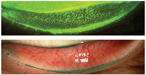
A unique photographic scale was found in this study to reliably grade lid wiper epitheliopathy. Photo: Jalaiah Varikooty, Centre for Contact Lens Research. Click image to enlarge. |
While the Korb scale has been routinely used to assess lid wiper epitheliopathy (LWE), no grading scale is perfect. A new study sought to develop an updated kind of photographic scale to grade the condition, and it worked just as well as the Korb staging, with some additional advantages.
As well as its development, the study also assessed validation and graders’ preference for using a photographic scale for LWE. Named the Photographic Lid Wiper Epitheliopathy (PLWE) scale, it was developed by taking LWE images from 57 screened patients confirmed to have the condition in both eyes. Validating the scale was accomplished by including a 20-image set displaying varying degrees of epitheliopathy, ranging from none to severe. Finally, grading validity and reliability were tested through observers’ grading assessments of the selected images using the PLWE scale and separately, in another session, grading using the Korb scale.
Results were near universal in preferring the visual-based approach, with 95% of graders claiming the PLWE scale was easier to use than Korb. The same percentage additionally would consider using the PLWE scale in clinical practice. Reliability of both scales were comparable at greater than 0.1.
Based on this reliability, the authors of the study believe clinicians would be better suited using a 0.5 grading step to better monitor changes using the PLWE scale. What’s more, they advise extrapolation of grading estimates beyond the limit illustrated in the PLWE if epitheliopathy seems greater than a 3.0 grade.
The study wanted to look at any discrepancies when comparing the two scales, and most of this came from the middle of the range, where the Korb scale tended to underestimat the amount of LWE. Conversely, the scales exhibited greatest agreement toward the extreme ends, indicating that graders can accurately assess a lack of epitheliopathy and large extents of it.
The Korb protocol specifically indicated clinicians’ underestimation of LWE at greater severity levels, and the authors believe this is partially due to encouraged usage of a 0.5 increment when using the PLWE scale, including extrapolation to 3.5.
Potentially making this scale easier to use is the reliance on a reference set of photos, obviating the Korb scale’s requirement to mentally measure length and width of curvilinear ocular anatomy. This way, graders don’t have to mentally subtract the line of Marx, leaving images to be evaluated in their easier, natural state.
In terms of clinical application, the authors believe their scale to help in assessing LWE in association with dry eye. In their publication of their results, they posit that “the linear design of the PLWE affords an appropriate level of discrimination for the clinician to aptly determine LWE severity.” Adding to its advantage is the fact that “visual scales, like this one, are simple to administer and require little to no instruction,” offering clinicians a more efficient tool.
Lievens CW, Norgett Y, Allen PM, Vianya-Estopa M. Development and validation of a new photographic scale to grade lid wiper epitheliopathy. Cont Lens and Int Eye. 2022. [Epub ahead of print]. |
