 |
| A decrease of macular RNFL thickness appeared more often in this study in patients with both glaucoma and high myopia than in high myopes alone, arguing for its diagnostic value. Photo: Jarett Mazzarella, OD, and Justin Cole, OD. Click image to enlarge. |
With research showing that patients with high myopia were six times more likely to develop glaucoma, it’s important to understand the role played by refractive error in determining risk for open-angle glaucoma, especially with the prevalence of both diseases growing substantially. In a new study, researchers evaluated the differences in structural parameters in patients with glaucoma, high myopia and both diseases concurrently.
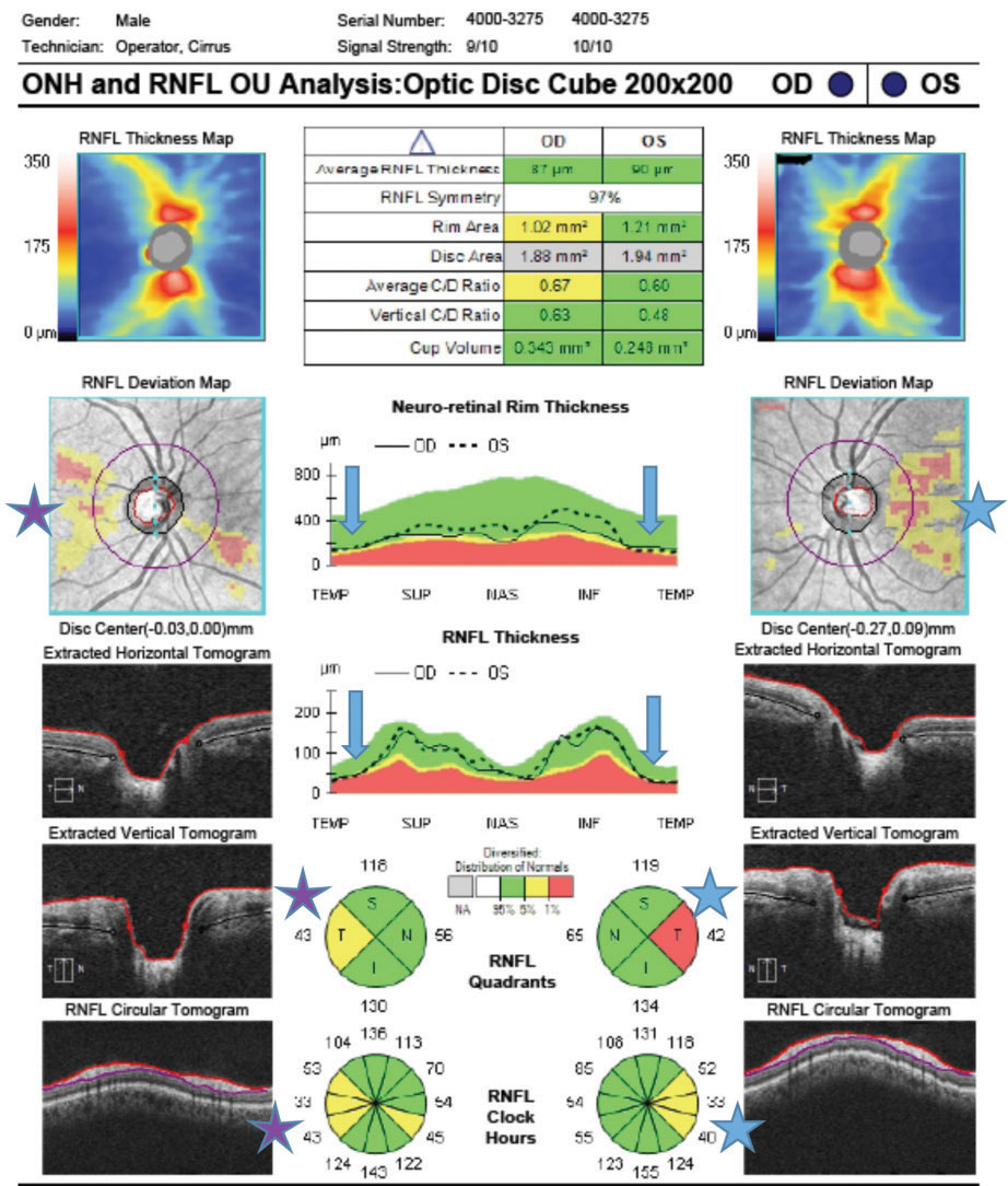
A total of 42 participants were included: 14 with OAG, 14 with myopia and 14 with both diseases. Mean peripapillary retinal nerve fiber layer (RNFL) thickness, RNFL in each quadrant, macular ganglion cell complex (GCC) and its layers, vessel density of optic nerve head and macula were all evaluated.
The myopia group showed the least thinning in the peripapillary RNFL thickness in the temporal quadrant and macular RNFL compared to other two groups. The highest macular vessel density in the inferior quadrant was in the myopia group in the superficial capillary plexus, deep capillary plexus and choriocapillaris. The myopia group showed highest density in the temporal quadrant and in total vessel density of optic nerve head at the superficial capillary plexus and at the deep capillary plexus.
The findings also showed that mean peripapillary RNFL thickness correlated positively with all the evaluated macular structural parameters. The strongest association was found between mean peripapillary RNFL thickness and macular GCC thickness in glaucoma patients and those with both conditions.
“In addition, we found that peripapillary RNFL thickness in the temporal quadrant was significantly higher in patients with high myopia” vs. the other two groups. “This corresponds with other studies’ results that observed thickening of RNFL in the temporal quadrant in high myopia due to the temporalization of RNFL due to increased axial length,” the authors explained in their article on the study. “The RNFL thickness in the temporal quadrant is expected to be the least affected by high myopia changes suggesting that a decrease in RNFL thickness in this part could indicate glaucomatous changes.
“This data suggests glaucomatous changes in persons with significant myopia may be best determined in part by evaluating the thickness of macular GCC and its layers,” the authors continued. They attribute this to the finding that only macular RNFL thickness was significantly lower in patients with both conditions compared with myopia-only patients. “This suggests that the decrease of macular RNFL thickness may be advantageous for diagnosing glaucoma in patients with high myopia.”
Markeviciute A, Januleviciene, Antman G, et al. Differences in structural parameters in patients with open-angle glaucoma, high myopia and both diseases concurrently. PLoS One. June 22, 2023. [Epub ahead of print.] |
