 |
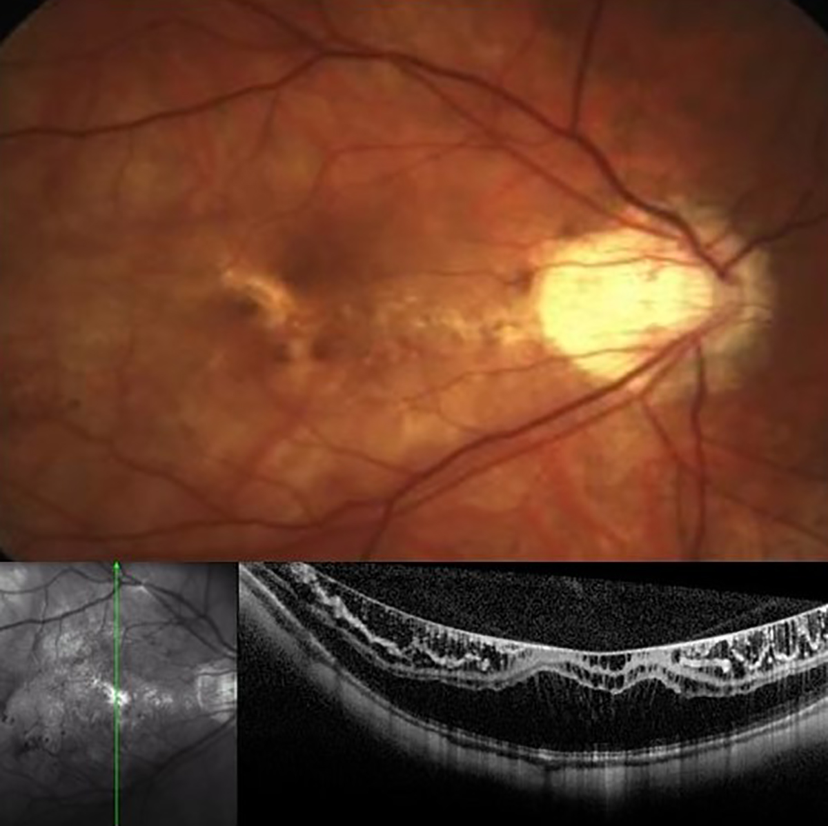
| The predominant pattern of advancement manifested in these pediatric myopes as the enlargement of diffuse atrophy, constituting more than half of the reported cases. Photo: Mohammad Rafieetary, OD. Click image to enlarge. |
Despite the significance of myopic maculopathy, there is limited documentation regarding its progression in children and adolescents. Its progression pattern in this specific demographic and the associated factors remain unclear, and knowing that would be helpful in timely intervention in individuals at risk of developing pathologic lesions in the fundus. Researchers in China investigated the progression of myopic maculopathy in children and adolescents with high myopia over a four-year follow-up period. The proportion of myopic maculopathy progression in this population was 12.2%. Enlargement of diffuse atrophy was the most frequent progressive change. Their findings were published in JAMA Ophthalmology.1
This hospital-based observational study with four-year follow-up included a total of 548 high myopic eyes (spherical power −6.00D or less) of 274 participants aged seven to 17 years (50.4% girls, mean age: 13.6 years). The mean axial length (AL) was 27.08mm and mean spherical equivalent was -9.12D.
The four-year progression of myopic maculopathy was found in 67 of 548 eyes (12.2%) of 274 participants with 88 lesion changes, including new signs of the tessellated fundus in 16 eyes (18.2%), diffuse atrophy in 12 eyes (13.6%), patchy atrophy in two eyes (2.3%), lacquer cracks in nine eyes (10.2%) and enlargement of diffuse atrophy in 49 eyes (55.7%).
Multivariable analysis revealed that worse best-corrected visual acuity (odds ratio; OR: 6.68), longer AL (OR: 1.73), faster AL elongation (OR: 302.83) and more severe myopic maculopathy at baseline (diffuse atrophy, OR: 4.52 and patchy atrophy, OR: 3.82) were associated with myopic maculopathy progression.
The same analysis noted that eyes categorized as myopic maculopathy C1 and C3 at the initial visit were at higher risk of myopic maculopathy progression than eyes with a normal fundus. “This suggests that, compared with adults, children and adolescents have a greater likelihood of progressing myopic maculopathy whenever a tessellated fundus was present,” the researchers wrote in their paper. “Therefore, it is imperative to closely monitor and implement proactive interventions for young individuals who just have mild fundus lesions and are likely to deteriorate further.”
A commentary on the study was also published in JAMA Ophthalmology and noted how the research addresses this current gap in knowledge about myopic maculopathy, shedding light on its progression during childhood and adolescence—crucial periods marked by advanced increase of the ocular axial length.2
“Given the aforementioned findings, it is probable that children exhibiting diffuse atrophy, potentially peripapillary diffuse atrophy, are more predisposed to developing new lesions of myopic maculopathy and progressing toward a more severe form of the condition,” the commentary author wrote.
The commentary also found it noteworthy that 12 eyes developed diffuse atrophy in individuals who exhibited no signs of such atrophy at baseline, including those with a normal fundus or tessellation alone. “This suggests that relying solely on a normal fundus or tessellated fundus at baseline is not sufficient for reassurance,” the author proposed. “It underscores the necessity for ongoing, regular and long-term monitoring to detect and track the development and progression of myopic maculopathy.”
She added that the integration of advanced imaging modalities such as ultra-widefield OCT could enhance the ability to forecast the risk of myopic maculopathy progression and future visual impairment in individual children, perhaps even better than fundus photography.2
1. Jian F, Wang D, Xiao O, et al. Four-year progression of myopic maculopathy in children and adolescents with high myopia. JAMA Ophthalmol. January 25, 2024. [Epub ahead of print]. 2. Ohno-Matsui K. Insights into childhood myopic maculopathy. JAMA Ophthalmol. January 25, 2024. [Epub ahead of print]. |
