 |
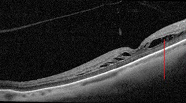
Foveoschisis in a high myope similar to those studied. EMSSRD gives the appearance of a macular hole. Photo: Sara Weidmayer, OD. Click image to enlarge. |
Researchers recently identified a new clinical entity in myopic eyes that they termed “extreme macular schisis simulating retinal detachment” (EMSSRD). They observed this finding on high-resolution OCT after closely evaluating patients who at first appeared to have macular hole retinal detachments.
“EMSSRDs resemble a macular hole retinal detachment, but they differ by having thin remnants of the retina on the retinal pigment epithelium,” the researchers explained in their paper.
To further characterize this finding, they analyzed data of 617 highly myopic eyes with myopic tractional maculopathy. They diagnosed EMSSRD on OCT based on high retinal elevation (>500µm), less obvious columnar structures and presence of thin remnants of outer retinal tissues above the retinal pigment epithelium.
Of the total eyes, 4% had EMSSRD. All of these eyes had macular atrophy caused by myopic macular neovascularization. The researchers noted that less than 1% of eyes progressed to macular hole retinal detachment, and these detachments all began away from rather than within the macular atrophy.
“This suggested that although the tissue adhesion was strong in the area of the macular neovascularization-related macular atrophy, the retinal detachment can spread toward the area of macular atrophy with a strong adhesion because of continued tractional forces,” the researchers wrote in their paper. “When the tractional force exceeded adhesion, it resulted in a macular hole retinal detachment.”
Almost 2% of eyes required vitreoretinal surgery, but the researchers found no significant difference between pre- and post-op best-corrected visual acuity in eyes operated on due to worsening EMSSRD or progression to macular hole retinal detachment. There was no significant difference in best-corrected visual acuity improvement between the two types either.
“The differentiation between EMSSRD and macular hole retinal detachment is important because the treatment strategies including urgent vitrectomy for macular hole retinal detachment are different from that of EMSSRD,” they concluded.
Uramoto K, Azuma T, Watanabe T, et al. Extreme macular schisis simulating retinal detachment in eyes with pathologic myopia. Retina. May 27, 2022. [Epub ahead of print]. |
