 |
A 34-year woman presented complaining of a painful upper eyelid OD of two days’ duration following application of false eyelashes. She had removed the lashes the day before and explained that her lid remained swollen, “purple” and painful. She denied trauma, systemic disease or allergies of any kind.
Clinical Findings
Her best-corrected entering visual acuities were 20/20 in the right eye and 20/20 in the left at distance and near. Her external examination was remarkable for a painful and swollen right upper eyelid, tender to the touch, which is demonstrated in the photograph below on the left. Her extraocular motilities were full, her confrontation visual fields were intact and there was no evidence of afferent pupillary defect. Her posterior segment findings were normal and Goldmann applanation tonometry measured 17mm Hg OU.
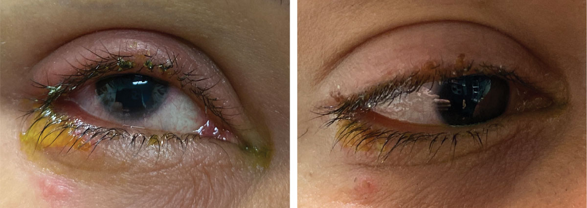 |
The patient’s initial presentation is shown at left and her appearance after resolution at right. What do you see here? Click image to enlarge. |
Additional Testing
The patient was further assessed by palpation of the injured area and inspection of the tissue for firmness, intactness (puncture wound), bleeding or suppurative oozing. The lashes were inspected for lingering “glue.” The fornix regions were evaluated for foreign material. The cornea was inspected for superficial injury.
Two for One
The diagnosis in this issue is a combination of contact dermatitis with a mild preseptal cellulitis. Below, we will discuss each in turn.
Contact Dermatitis
This is an inflammatory reaction of the eyelid(s) and/or periocular adnexa. The presentation may be unilateral or bilateral, depending upon the exposure. In acute cases, the patient may present with generalized lid erythema and edema, as well as pruritic vesicles or bullae.1 Chronic cases may show eczema, a characteristic thickening and scaling of the involved skin which is typically seen in the atopic variant (atopic dermatitis).2
Symptoms include intense itching and burning of the skin and eyes and associated tearing (lacrimation). The palpebral and bulbar conjunctiva may also be involved, demonstrating variable injection and chemosis. Palpebral follicles may be seen in severe cases. Corneal involvement is rare, though chronic eye rubbing may lead to punctate keratopathy or epithelial erosion. Vision is not typically affected to any substantial degree, and preauricular lymphadenopathy is characteristically absent.
The history is of paramount importance in correctly diagnosing contact dermatitis. Many cosmetics, cleaning agents, fabrics, fragrances, medications, nickel and even metallic jewelry can be implicated in this skin reaction.2-4 Typically, since the reaction connotes some acute exposure to an offending allergen, it is assumed that the patient recently encountered something “new.” However, many agents implicated in contact dermatitis represent weak sensitizers, and the exposure may occur over weeks, months or even years.4 In cases involving the ocular tissues, one should consider those substances which are readily applied or coincidently touch the skin around the eyes, particularly makeup (eyeliner, eye shadow, mascara) or makeup remover, sunscreen, contact lens solutions or metal spectacle frames. A wide range of ophthalmic medications may also have the capacity to induce allergic contact dermatitis.5
Contact dermatitis may be subdivided into several categories: irritant contact dermatitis, allergic contact dermatitis and photoallergic contact dermatitis.
Irritant contact dermatitis (ICD) accounts for nearly 80% of all cases seen clinically.1 The condition, which is also referred to as non-allergic contact dermatitis, represents a non-preprogrammed, non-immunologic, local inflammatory reaction to a chemical or physical irritant. This may include substances such as soaps or detergents, solvents (e.g., paint thinner, acetone), or particulate matter such as fiberglass insulation. Typically, the severity of the ICD reaction is proportionate to the amount and duration of irritant exposure. The mechanism of action involves a direct, local cytotoxic effect on the epidermis, leading to subsequent keratinocyte damage.6 ICD can affect any individual, regardless of their immune status. A well-known example of ICD involves exposure to the plant Toxicodendron radicans, better known as poison ivy. Atopic individuals are especially susceptible to ICD and may have a lower threshold for irritant exposure.
Allergic contact dermatitis (ACD), by its very definition, affects only individuals who are genetically preprogrammed toward allergic hypersensitivity. Like all allergic reactions, ACD occurs in two phases: a sensitization process, in which specific antibodies are generated toward the allergen and an elicitation phase in which the actual cellular response occurs. The mechanism of ACD involves a delayed, or Type IV, hypersensitivity reaction.6 This is a cell-mediated response, which employs discrete subpopulations of T-lymphocytes and immunoregulatory cytokines, including tumor necrosis factor alpha and interleukins 1, 13 and 18.7 Mast cells, which play a pivotal role in immediate Type I hypersensitivity reactions such as allergic conjunctivitis are not central to Type IV reactions like ACD. However, the mast cell still has an important function, as it indirectly helps to control neutrophil recruitment.8
Photoallergic contact dermatitis (PACD) is an eczematous skin reaction initiated by an otherwise benign substance on the skin, which becomes noxious when exposed to ultraviolet light. We refer to substances that induce PACD as photosensitizing agents. PACD characteristically occurs only with areas of exposed skin, such as the hands and face. Known photosensitizing agents include tar-based products, octyl dimethyl PABA (an ingredient in some commercial sunscreen formulas), certain forms of vegetation (including carrot, lemon and mustard plants) some oral antibiotics such as tetracycline and topical NSAID medications such as ketoprofen, diclofenac and indomethacin.9,10
In cases of contact dermatitis, attempts should be made to identify the causative agent. Often, this can be accomplished with a careful history. In cases where this is not clear, epicutaneous patch testing can isolate the offending substance. Patch testing involves standardized samples of known allergens, placed on small delivery vehicles and applied to the skin of the upper back for two days.11,12 The test is typically performed only by an experienced dermatologist.
Once identified, the ideal long-term solution is to remove or avoid the causative agent. Short-term management involves emollients, treatment of secondary infection (if present) and downregulation of the immune response. The easiest method of arresting and reversing the out-spilling of chemical modulators is the cold compress. Here, vasoconstriction slows the movement of the chemokines, cytokines and cellular immune system, which create the immune reaction and perpetuate tissue edema.
Topical corticosteroids are the first medicinal line of therapy for most individuals, since these agents directly suppress the recruitment of polymorphonuclear leucocytes and reverse capillary permeability.12 Low-potency steroids such as hydrocortisone and desonide may be safer for use on the face, though stronger steroids such as triamcinolone, clobetasol proprionate or betamethasone dipropionate can be employed for moderate to severe disease.13 Steroid creams or lotions used twice daily for 10 to 14 days are usually very effective. Care must be taken to apply these preparations only to the skin of the lids and ocular adnexa, as they are not appropriate for use in the eye. Fluorometholone ointment or antibiotic/steroid combination ointment are often substituted by eye care practitioners as they may have samples to provide.
Ironically, some topical steroids themselves be allergenic, confounding management.14 In these cases calcineurin inhibitors such as tacrolimus and pimecrolimus may be useful.12
More severe cases may require systemic corticosteroids; oral prednisone (0.5-1mg/kg/d for two to three days, and tapered over one to two weeks) is the preferred course of therapy for recalcitrant contact dermatitis.1,6
The use of antihistamines is somewhat controversial in managing contact dermatitis. Since histamine release from mast cells is not central to the pathophysiology of the disorder, antihistamines would seem to be superfluous. However, oral agents such as cetirizine 10mg or desloratadine 5mg once daily may help to curtail the itching to some extent, and hence may be beneficial in addition to topical corticosteroid therapy.
When the conjunctiva is involved supportive therapy is beneficial. This includes cold compresses and the liberal use of ocular lubricants. Topical antihistamine/mast cell stabilizer products (e.g., OTC Pataday or Zaditor once daily in the affected eye as needed) may provide palliative relief of conjunctival itching, as well as ocular swelling and hyperemia. More severe involvement may warrant topical ophthalmic corticosteroids, such as 1% prednisolone acetate or 0.5% loteprednol etabonate every two to four hours for several days.
Preseptal Cellulitis
It is presumed in this case, because the eyelid was warm and firm to the touch with “purplish” color, that eye rubbing created a skin breach permitting the inception of infection creating a mild preseptal cellulitis (PC).
PC is an infection within the eye lid anterior to the orbital septum.1-5 Signs and symptoms include variable pain upon palpation, redness, swelling and “red-purple” skin coloration that is firm and warm to the touch.1-5 Other ocular signs include conjunctival injection, edema and depending upon the extent and severity of the periorbital processes, corneal insult and in rare instances limited ocular motility.5-7 Eyelid infections involving the orbit and adnexa have been organized via the modified Chandler classification into two forms: preseptal (Stage I-Preseptal cellulitis, II-orbital cellulitis, anterior to the orbital septum) and retroseptal (Stages III-Subperiosteal abscess, IV-Orbital abscess, V-Cavernous sinus thrombosis) posterior to the orbital septum.8,9
The etiologies of preseptal cellulitis include untreated hordeolum, dacryocystitis, sinusitis, eyelid trauma, eyelid infection secondary to puncture wound (foreign body, insect bite or sting) and infection by way of communication with a sinus following orbital fracture.1-13 Microbiologic cultures identify the most common pathogen as Staphylococcus aureus.9-12 There is no predilection for gender, age, or region. Immunosuppression may increase the risk.6
Preseptal cellulitis begins when inoculating microbes seed infection in the affected region. This can occur secondary to acute dacryocystitis, chronic sinusitis/upper respiratory infection, puncture wound from a foreign body from blunt or projectile trauma, an insect bite or sting, or as a result of chronic hordeola or chalazia.10,14 Iatrogenic causes include dacryocystorhinostomy, nasolacrimal probing, nasolacrimal stenting, surgical reduction of orbital or eyelid abscess, chalazion and cilia epilation.10,14,15 The most common microorganisms recovered include Staphylococcus(including methicillin-resistant Staphylococcus aureus, MRSA) and Streptococcus species, followed by Haemophilus influenzae and Klebsiella pneumonia.14-17
PC is particularly dangerous because the vessels of the face and orbit are well connected, with an interdigitating vascular web.18-20 Directly or indirectly the orbital venous system via the cavernous sinus and because there is no valve system restricting the direction of blood flow has access to the cranium.18-20
In order for the lids to maintain functional movement, rigid anatomical landmarks must provide shape and stability.21,22 The tarsal plates are found in both the upper and lower eyelids extending across the width the globe maintaining a contoured margin to track with the eye’s curvature.21 The muscles that help to elevate the lid are the levator palpebrae superioris and the muscle of Müller.
The muscular portion of the levator terminates superiorly in a broad flat tendon known as the levator aproneurosis.21 The tendon runs the entire width of the eyelid, inserting into the tarsus and the connective tissue that surrounds it.21 There is fibrous connective tissue between the Müller’s muscle and the palpebral conjunctiva, creating a natural barrier.22
The pretarsal portion of the orbicularis oculi is situated within the lid anterior to the tarsal plates.21 The orbital septum is a connective tissue sheet that forms a barrier between the orbital contents and orbital fat and the eyelid.21 It extends circumferentially around the entire orbital rim, inserting into the tarsal plate connective tissue.18,21 The only breaks in the orbital septum occur where vessels and nerves splice through it on their way to anterior structures and where the levator aponeurosis passes through it to insert on the connective tissue of the tarsal plates and dermis.18,21
The anatomy and blood supply of this region make it an ideal habitat for a pocket of infection to prosper and potentially spread into the orbit, cavernous sinus, blood or brain.1-25
Preseptal cellulitis can be conservatively managed with hot compresses at the site of infection to stimulate the body’s immune repose to the local region as well as broad spectrum oral antibiotics and/or topical antibiotics if penetration to deeper levels of the tissue is perceived to be necessary and oral analgesics.1-17,26-31
When lesions are small, focal, superficial and painful, they can be decompressed and allowed to passively drain by creating an opening with a small-gauge needle; epilating an obstructing eyelash or opening a visibly blocked gland.30,31The skin over the lesion can be anesthetized with topical anesthetic to aide in the comfort of the procedure; however, injectable anesthetic is rarely used as adding additional volume to an already congested region inhibits the agent’s diffusion.32
The oral antibiotic classes that are commonly used include the penicillins (cloxacillin, dicloxacillin, flucloxacillin) 250-500mg BID-QID, the cephalosporins (cephalexin, cefadroxil, cephradine) 250-500mg BID-QID, the macrolides (azithromycin as directed on Z-Pak, clarithromycin 500mg BID) and fluoroquinolones (ciprofloxacin, levofloxacin) 500mg BID-QID.26-28
Topical and oral antibiotics should never be tapered, and the duration should be seven to 10 days depending upon the severity of the infection or the area involved.
In more severe cases or cases with a larger area of infection, intravenous antibiotics can be initiated. In cases of concurrent dacryocystitis, epiphora may result, leading to a lateral canthus fissure or other ulcerative defects secondary to the drying effects of the sodium laden tears.33 In these cases a topical antibiotic ointment can augment a skin moisturizer to protect against infection and aid in lesion resolution.
Management Approach
We used topical anesthetic on a cotton-tipped applicator as a solvent to loosen the remaining “false eyelash” glue; when it was loose, we removed it with forceps. Once the lashes were free from adhesive agent, we asked the patient to use topical OTC hydrocortisone cream TID with cold compresses. We also initiated 500mg cephalexin, PO BID for seven days and arranged a follow up appointment to check progress in three days. The 90% three-day resolution photograph is included.
It is unclear why she would react this way in only one eye. It is possible she was allergic in both eyes but initiated a skin breach OD during eyelid rubbing, or perhaps there was an irregularity during the application procedure producing the concomitant preseptal infection. Using the principles of conservative management, a combination approach achieved resolution.
Dr. Gurwood is a professor of clinical sciences at The Eye Institute of the Pennsylvania College of Optometry at Salus University. He is a co-chief of Primary Care Suite 3. He is attending medical staff in the department of ophthalmology at Albert Einstein Medical Center, Philadelphia. He has no financial interests to disclose.
|
1. 2. Horner, F. Uber eine form von ptosis. Klin Monatsbl Augenh 1869;7(1):193. 3. Tantum, LA. Pupil anomalies. In: Onofrey BE, ed. Clinical optometric pharmacology and therapeutics. Philadelphia, PA.; J.B. Lippincott Co. 1991;13:1-13. 4. Burde, RM, Savino, RJ, Trobe, JD. Anisocoria and abnormal pupillary light reaction. In: Burde, RM, Savino, PJ, Trobe, JD, eds. Clinical decisions in neuro-ophthalmology, 2nd ed. St. Louis, MO.; Mosby Year Book Inc. 1992:321-346. 5. Myles, WM, Maxner, CE. Localizing value of concurrent sixth nerve paresis and postganglionic horners’s syndrome. Can J Ophthalmol 1994;29 91 0:39-42. 6. Maloney, WF, Younge, BR, Moyer, NJ. Evaluation of the causes and accuracy of pharmacologic localization in horner’s syndrome. Am J Ophthalmol 1980;90:394-402. 7. Bates, AT, Chamberlain, S, Champion, M, et al. Pholedrine–a substitute for hydroxyamphetamine as a diagnosis eye drop test in Horner’s syndrome. J Neurology, Neurosugery, and Psychiatry 1995;58:215-217. 8. Thompson, HS, Pilley, SFJ. Unequal pupils- a flow chart for sorting out the anisocorias. Survey Ophthalmol 1976;21(1):45-48. 9. Cullom, RD, Chang, B. Neuro-ophthalmology : Horner’s Syndrome. In: Cullom, RD, Chang, B, eds. The Wills Eye Manual, 2nd ed. Philadelphia, PA.; J.B. Lippincott Co. 1993: 241-246. 10. Sartori, F, Rea, F, Calabro, F, et al. Carcinoma of the superior pulmonary sulcus. J ThoracCardiovasc Surg 1992;104:679-683. 11. Majeed A, Ribeiro NP, Ali A, et al. A rare presentation of spontaneous internal carotid artery dissection with Horner's syndrome, VIIth, Xth and XIIth nerve palsies. 12. Alonso Formento JE, Fernández Reyes JL, Envid Lázaro BM, et al. Horner's syndrome due to a spontaneous internal carotid artery dissection after deep sea scuba diving. Case Rep Neurol Med. 2016;2016:5162869. 13. Macdonald DJ, McKillop EC. Carotid artery dissection after treadmill running. Br J Sports Med. 2006;40(4):e10; discussion e10. 14. Verdin V, Holemans C, Otto B, et al. Horner's syndrome revealing a spontaneous carotid artery dissection. Rev Med Liege. 2013;68(1):11-5. 15. Shankar Kikkeri N, Nagarajan E, Sakuru RC, Bollu PC. Horner Syndrome Due to Spontaneous Internal Carotid Artery Dissection. Cureus. 2018;10(9):e3382. 16. Chan CC, Paine M, O'Day J. Carotid dissection: a common cause of Horner's syndrome. Clin Exp Ophthalmol. 2001;29(6):411-5. 17. Schelfaut D, Dhondt E, De Raedt S, et al. Carotid artery dissection: three cases and a review of the literature. Eur J Emerg Med. 2012;19(3):181-7. 18. Solomon S, Lustig JP. Benign Raeder's syndrome is probably a manifestation of carotid artery disease. Cephalalgia. 2001;21(1):1-11. 19. Zournas C, Kapaki E, Doris S, et al. Raeder's syndrome. Report of two cases. Int Angiol. 1995;14(4):415-7. |
