 |  |
Vital dyes are often used in surgical procedures to enhance the visibility of targeted tissues. Some vital dyes used by ophthalmologists include trypan blue, sodium fluorescein, indocyanine green and gentian violet. Of these, trypan blue is the most frequently used in cataract surgery due to its safety, availability and effectiveness.1 Trypan blue has been used in cataract surgery since the late 1990s to stain the anterior capsule and improve visibility for the surgeon.1
Visibility is Vital
The creation of the capsulorhexis in cataract surgery is a delicate procedure that requires careful removal of a circular portion of the anterior lens capsule. This process is far more difficult without visibility of the red reflex and anterior capsule. Poor visibility can increase the risk of radial tears, capsular rupture and intraocular lens (IOL) displacement.1 Visualization can be facilitated by dimming the operating room lights, using high magnification in the microscope and oblique illumination or applying a high-density viscoelastic such as sodium hyaluronate.2
But for mature, traumatic and pediatric cataracts, these techniques are often not enough, and a quick and easy injection of trypan blue is necessary, significantly increasing the success rates of capsulorhexis procedures.1 This vital dye provides uniform staining of the anterior capsule in 40 to 60 seconds and can help highlight any existing capsular tears.1 Patients with corneal opacities, abrasions, edema or arcus senilis may also benefit from the use of trypan blue during cataract surgery.1
Steps for Staining
Various techniques exist for staining the capsule with trypan blue. Surgeons can inject the dye into the anterior chamber under an air bubble or viscoelastic agent or mix trypan blue with a viscoelastic agent and then inject the mixture under an air bubble.1
Some surgeons use an even simpler technique: intracamerally injecting trypan blue through the side-port incision following an injection of non-preserved lidocaine (Figure 1). The dye remains in the eye for 40 to 60 seconds and is then removed by the injection of a viscoelastic agent into the anterior chamber. As a result, the capsule is stained blue and ready for the capsulorhexis procedure.
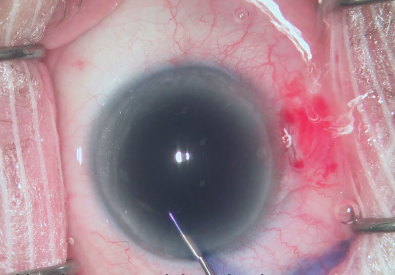 |
| Fig. 1. The surgeon injects the vital dye through the side-port incision to increase visibility during cataract surgery. Click image to enlarge. |
Pros and Cons
The application of trypan blue has its advantages, but it’s not without the potential for adverse reactions, for which comanaging clinicians should be prepared. Trypan blue has a risk, albeit minimal, of toxicity and spikes in IOP.1 Rarely, it can cause lens epithelial cell death, which can actually reduce the incidence of posterior capsule opacification.2 Other studies show that trypan blue can discolor high water content hydrogen IOL implants and inadvertently stain the posterior lens capsule and anterior face of the vitreous.1 The dye can lead to a decrease in lens capsule elasticity and increase the risk of capsular tears.3
Optometrists should note whether a patient had an instillation of trypan blue during surgery and check for any adverse reactions during the early post-op follow-up period.
Ms. Tran is a fourth-year student at Pacific University College of Optometry.
Dr. Skorin is a consultant in the Department of Surgery, Community Division of Ophthalmology at
the Mayo Clinic Health System in Albert Lea, MN.
| 1. Jhanji V, Chan E, Das S, et al. Trypan blue dye for anterior segment surgeries. Eye (Lond). 2011;25(9):1113-20. 2. Olson RJ, Jin GJC, Ahmed IK, et al. Cataract surgery from routine to complex. Thorofare: Slack; 2011:76. 3. Jacobs DS, Cox TA, Wagoner MD, et al. Capsule staining as an adjunct to cataract surgery. Ophthalmology. 2006;113(4):707- 13. |
