 A 22-year-old black female presented for an eye exam to determine if she met the visual requirements for obtaining a driver’s license in the state of Florida. She reported being very nearsighted, and that she had poor vision her entire life. The patient also said that she wore glasses for as long as she could remember, and had undergone multiple eye examinations since early childhood.
| |
|
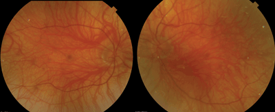
1. Our patient’s fundus evaluation reveals significant changes in both eyes (OD left, OS right).
|
Her medical history was significant for relatively light skin pigmentation, compared to her immediate family members.
Her visual acuity measured 20/80 OU with a spectacle correction of -14.50 + 2.75D X 110 OD and -13.50 +1.50D X 080 OS. At near, she was 20/40 OU. Extraocular motility testing revealed horizontal nystagmus in all positions of gaze. She did not display a head turn, nor did she have a noticeable null point. Her pupils were equally round and reactive, with no evidence of afferent defect OU.
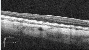
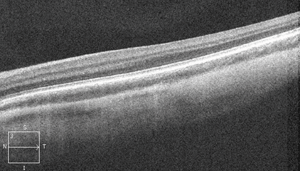
Dilated fundus exam revealed pronounced changes (figure 1). We also obtained a spectral-domain optical coherence tomography (SD-OCT) scan (figures 2 and 3).
Take the Retina Quiz
1. Based on the data provided, what changes would you expect to see in the anterior segment?
a. Keratoconus.
b. Anterior uveitis.
c. Iris transillumination defects.
d. Posterior subcapsular cataract.
2. How would you characterize the SD-OCT finding in our patient?
a. Macular edema.
b. Choroidal neovascularization.
c. Poorly differentiated macula.
d. Improper scan alignment.
3. What is the likely diagnosis?
a. Oculocutaneous albinism.
b. Myopic degeneration.
c. Congenital nystagmus.
d. Choroideremia.
4. What is the cause of her poor vision?
a. Macular edema.
b. Macular hypoplasia.
c. Optic nerve hypoplasia.
d. Choroidal neovascularization.
5. Does our patient meet the usual legal requirements for obtaining a driver’s license?
a. Yes.
b. No.
c. Yes––for daytime driving only.
d. Yes––with low vision aids.
Discussion
From external appearance alone, it was obvious that our patient had cutaneous albinism. And, given her history of poor vision and nystagmus, it was readily apparent that the albinism also had affected her eyes.
On slit-lamp evaluation, she had iris transillumination defects, and on dilated fundus exam we noted a striking lack of pigmentation within the retinal pigment epithelium. All these findings are consistent with oculocutaneous albinism.
Albinism represents a hereditary group of disorders characterized by a congenital lack of melanin pigment.1 Although the condition may be isolated to the eye (ocular albinism), most cases also involve the skin and hair––as was the case in our patient. Often, these individuals present with an obvious lack of skin pigmentation, their hair has a bleached white appearance, and their irides are light blue or almost white in appearance.
|
|
|
|
|
2, 3. How do you explain her SD-OCT findings (OD top, OS bottom)?
|
There are four types of oculocutaneous albinism:
• Type 1 is characterized by white hair, very pale skin and lightly colored irides.
• Type 2 is often less severe, and the individual’s skin usually is a creamy white color. Also, the patient’s hair may be light yellow, blond or light brown.
• Type 3 includes a variant called rufous oculocutaneous albinism, which usually manifests in dark-skinned people. Affected individuals often have ruddy-brown skin, ginger-tinted or red hair, and hazel or brown irides. This form is often associated with milder vision abnormalities than the other forms of oculocutaneous albinism.
• Type 4 exhibits signs and symptoms similar to those associated with type 2.
Because most of these clinical features overlap (e.g., those produced by types 2 and 4), the four forms of oculocutaneous albinism are most accurately distinguished by their underlying genetic cause––although all types of the condition are autosomal recessive.1,2
Despite clinical variations between the types of albinism, most patients have varying degrees of nystagmus and reduced visual acuity. This is due to the lack of pigmentation within the fundus as well as macular hypoplasia––the most consistent feature of albinism.1
The macula and fovea become poorly differentiated, with more rods and fewer cones present in the fovea. Also, the foveal pit may be reduced or not even present. This is evident in our patient on the SD-OCT scan, where the normal foveal depression is absent. Interestingly, the junction of the inner and outer photoreceptor segments can still be seen on the scan. Perhaps that finding explains why our patient’s acuity is 20/80, while most macular hypoplasia patients often see 20/100 to 20/200.
Iris transillumination defects are another common feature of albinism, and can easily be seen with minimal light directed at the eye. These patients are often highly photophobic.
Our patient didn’t know what type of oculocutaneous albinism she had. Based solely on external appearances, however, we were fairly certain that it was type 3.
Finally, to address the question as to whether her acuity sufficiently meets the legal requirements to obtain a driver’s license… The answer is no. In Florida, the law states that the driver’s best-corrected visual acuity must be at least 20/70 in each eye; or if one eye is worse than 20/200, the fellow eye must be 20/40 or better.3 Thus, 20/80 OU fails to meet that mandate.
We explained these findings to our patient. Afterward, we provided her with several forms to fill out, so she could obtain disability as well as any rights and privileges offered to legally blind individuals.
Answers: 1) c; 2) c; 3) a; 4) b; 5) b.
1. Russell-Eggitt IM. Albinism. Pediatric Ophthalmology and Strabismus, 4th ed. Edingburgh: Saunders Elsevier; 2014:393-9.
2. US National Library of Medicine. Genetics Home Reference: Oculocutaneous Albinism. Available at: http://ghr.nlm.nih.gov/condition/oculocutaneous-albinism. Accessed June 12, 2014.
3. Florida Department of Highway Safety and Motor Vehicles. Report of Eye Exam. Available at: www.flhsmv.gov/hsmvdocs/vision.pdf. Accessed June 12, 2014.
