 |
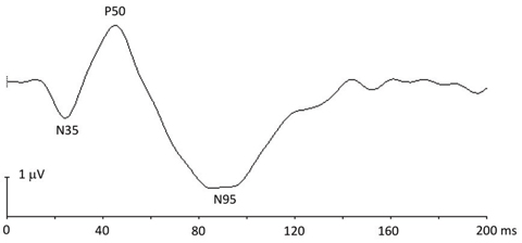
| Abnormalities shown on pERG were able to accurately predict RNFL rim thinning in this study. Photo: Hua Bi, OD, PhD. Click image to enlarge. |
Researchers based in New York recently examined the relationship between pattern electroretinogram (pERG) and OCT-derived optic nerve head (ONH) measurements, after controlling for disc area. They found significant associations between pERG parameters and ONH morphology measurements such as rim area and Bruch’s membrane opening-minimum rim width (BMO-MRW) sectors.
The study assessed 32 eyes of 20 subjects with preperimetric glaucoma. pERG parameters (Magnitude, MagnitudeD and MagnitudeD/Magnitude) and ONH measurements were analyzed after controlling for disc area. Magnitude and MagnitudeD were significantly associated with rim area. All pERG parameters significantly correlated with BMO-MWR sectors—temporal superior and nasal inferior—as well as with RNFL sectors—superior, nasal superior and inferior.
“These findings suggest that, along with a global deterioration of the neuroretinal rim and its significant thinning in preperimetric glaucoma, there is a sectoral morphological change in the neuroretinal rim in the form of reductions in the BMO-MRW thickness measurements,” the authors wrote.
The researchers’ findings propose a degenerative pattern in which morphologic change occurs to the disc with possible concurrent retinal ganglion cell (RGC) dysfunction and subsequent axonal damage, accompanied by delay of axonal transport. The MagnitudeD parameter in pERG represents phase delays and an opportunity to detect RGC dysfunction preceding cell death. At this stage, RGC damage is potentially reversible.
“When examining preperimetric glaucoma patients, we recommend use of steady-state pERG with OCT-derived ONH morphology measures while controlling for disc area to increase diagnostic accuracy of the devices and to circumvent underestimation or overestimation of the neuroretinal rim,” they concluded.
Tirsi A, Gliagias V, Moehringer J, et al. Pattern electroretinogram parameters are associated with optic nerve morphology in preperimetric glaucoma after adjusting for disc area. J Ophthalmol. October 13, 2021. [Epub ahead of print]. |
