History

A nine-year-old black male presented for a routine eye examination. His chief complaint was that he wanted to correct his “shifty eyes,” which were a source of teasing for classmates at school. His systemic history was unremarkable. Additionally, he had no established ocular history and reported no allergies.
Diagnostic Data
His best-corrected visual acuity measured 20/200 O.U. at distance and near. External examination was positive for searching nystagmus with no null point. There was no evidence of afferent pupillary defect. The anterior segment was normal. His intraocular pressure measured 14mm Hg O.U. The dilated fundus findings are illustrated in the photographs.
Your Diagnosis
How would you approach this case? Does this patient require any additional tests? What is your diagnosis? How would you manage this patient? What’s the likely prognosis?
Discussion
Additional tests might include a 100-hue color vision test or panel D-15 color vision test, Amsler grid testing, visual field testing to rule out functional deficits and fundus photography. Sodium fluorescein angiography would not be indicated unless there was a reason to suspect chroidal neovascularization (CNV).
The diagnosis in this case is coloboma of both optic nerve heads. An ocular coloboma is defined as an area in the fundus that lacks tissue secondary to incomplete closure of the fetal embryonic fissure.1-7 Optic nerve colobomata (those limited to the optic nerve) are described as part of the spectrum of large, abnormally appearing optic discs, where tissue is missing. They are usually located inferonasally and may be associated with other colobomata.
Optic nerve coloboma (ONC), in general, is a congenital condition that typically is discovered during a fundus examination at the patient’s first eye exam. It may present unilaterally or bilaterally and can mildly to severely affect visual function.2
In children, ONC generally exhibits any or all of the following signs: afferent pupillary defect, loss or decrease of color vision, strabismus, searching nystagmus and decreased visual acuity.1-8 Children who present with ONC and decreased visual acuity without frank strabismus should be evaluated for uncorrected refractive error. If a significant ametropia is absent and normal foveal anatomy is intact, occlusion therapy may be attempted to rehabilitate visual acuity.1,2
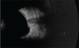
Fundus photograph and B-scan of our nine-year-old patient who presented with “shifty eyes.” What is your diagnosis?
The presence of decreased visual acuity varies greatly in cases of ONC and is dependant on the presence of damage to fibers which subserve the fovea.1,4 Strabismus is not present in all cases of ONC. When present, however, it is often secondary to poor vision or sensory deprivation of the involved eye along with associated foveal hypoplasia. When foveal hypoplasia is present it often includes the phenomenon of searching nystagmus.5
Afferent pupillary defects produced by this anomaly may be markedly pronounced, depending on the laterality and severity of the disc excavation.1-5 The extent of color vision impairment is also dependant on the amount of missing tissue.1-5
In many cases, ONC is accompanied by other ocular and/or systemic abnormalities.6,7 Colobomatous malformations may also include those of the fundus and lens.6,7 Retinal detachments are common in ONC patients and create an uncertainty as to the overall ocular and functional prognosis.6,7 ONC eyes are also susceptible to serous macular detachments.9,10 Serous fluid is derived from one of three places: fluid which perfuses into the retrobulbar space from surrounding orbital tissues, fluid from the peripapillary choriocapillaris or cerebrospinal fluid.9,10
ONC is a common finding in reno-ocular coloboma syndrome.5 These patients may present with colobomatous eye defects, renal hypoplasia, vesicoureteral reflux, high-frequency hearing loss and rarely, central nervous system abnormalities.6
There is no treatment for ONC. Newly-diagnosed patients must be examined for acro-reno-ocular syndrome as well as other systemic and ocular malformations. Patients should be closely monitored for retinal and/or macular detachment with routine fundus examinations and home Amsler grid testing.
Thanks to Virginia Donati, O.D., of Brampton, Ontario for contributing this case.
1. Cekić S, Stanković-Babić G, Visnjić Z, et al. Optic disc abnormalities - diagnosis, evolution and influence on visual acuity.Bosn J Basic Med Sci. 2010;10(2):125-32.
2. Kim MR, Park SE, Oh SY. Clinical feature analysis of congenital optic nerve abnormalities. Jpn J Ophthalmol. 2006;50(3):250-5.
3. Dutton GN. Congenital Disorders of the Optic Nerve: Excavations and Hypoplasia. Eye (Lond). 2004;18(11):1038-48.
4. Olsen TW, Summers CG, Knobloch WH. Predicting visual acuity in children with colobomas involving the optic nerve. J Pediatr Ophthalmol Strabismus. 1996;33(1):47-51.
5. Azuma N, Yamaguchi Y, Handa H, et al. Mutations of the PAX6 gene detected in patients with a variety of optic-nerve malformations. Am J Hum Genet. 2003;72(6):1565-70.
6 Schimmenti LA, Manligas GS, Sieving PA. Optic nerve dysplasia and renal insufficiency in a family with a novel PAX2 mutation, Arg115X: further ophthalmologic delineation of the renal-coloboma syndrome. Ophthalmic Genet. 2003;24(4):191-202.
7. Dureau P, Attie-Bitach T, Salomon R, et al. Renal coloboma syndrome. Ophthalmology. 2001;108(10):1912-6.
8. Olsen TW. Visual Acuity in children with colobomatous defects. Curr Opin Ophthalmol. 1997;8(3):63-7.
9. Perkins SL, Han DP, Gonder JR,et al. Dynamic atypical optic nerve coloboma associated with transient macular detachment. Trans Am Ophthalmol Soc. 2005(1);103:116-23.
10. Lin CC, Tso MO, Vygantas CM. Coloboma of optic nerve associated with serous maculopathy. A clinicopathologic correlative study. Arch Ophthalmol. 1984;102(11):1651-4.
