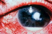
With improved technology, optometrists can now clearly see the retina through photography, which has become a routine screening tool in many offices. However, dilation and funduscopy are still necessary to diagnose and manage retinal disease.
As we see more patients who have systemic conditions (e.g., diabetes and hypertension) and age-related eye conditions, detection of early retinal changes is crucial. Here, Ill review the common techniques of posterior segment examination and provide recommendations to help you improve your observation of the retina.
Direct Ophthalmoscopy 101
The direct ophthalmoscope, which we commonly use to examine the retina, provides magnified views of retinal details, such as the optic disc, individual retinal vessels and the fovea. We can also use the direct ophthalmoscope to examine the crystalline lens and opacities of the posterior capsule. Dilation, while not necessary, can help maximize the usefulness of this technique.
Direct ophthalmoscopy is generally fast and easy to perform. Images appear upright and in normal orientation.
The disadvantages, however, include a limited and non-stereoscopic view of the fundus. This negates the advantages of a higher level of magnification.
The slit-lamp biomicroscope allows us to diagnose and monitor retinal disease in great detail. Several condensing lenses enable us to achieve the desired magnification. These lenses fall into two categories: non-contact lenses and fundus contact lenses.
Non-contact lenses, such as the 60D, 78D, 90D and Superfield NC lenses, provide a magnified stereoscopic view and an inverted, reversed image of the retina. The 60D lens provides the most magnification, while the 90D lens provides the least magnification but the largest visual field view. The 78D lens is a good all-purpose lens.
Newer non-contact lenses, such as the Digital Wide Field (Volk) and the Digital 1.0x imaging lens (Volk), may provide higher magnification without sacrificing much of the visual field view. Also, a non-contact ruby lens provides a view that is neither reversed nor inverted.
Use fundus contact lenses if you suspect subtle thickening or edema. They provide images at the same orientation as the retina. The Goldmann three- or four-mirror lenses and the contact ruby lens (figure 1) are commonly used fundus contact lenses.
 |
| Figure 1. An assortment of non-contact and contact condensing lenses. |
A well-dilated pupil is very important for obtaining an adequate view of the posterior segment with the slit-lamp biomicroscope. You can then use two techniques for viewing the posterior segment.
For gross scanning of retinal details to find large pathological lesions, use moderate illumination and a wider slit lamp beam. To adequately scan the fundus, move the microscope rather than the lens.
This technique can also help you assess the mid- and far periphery if the patient changes his or her gaze position. You can enhance the view in far eccentric gaze by reactively tilting the lens in your hand.
A second method of slit-lamp biomicroscopy includes evaluation of fine details, retinal thickness and/or any edema that may be present. For this evaluation, use a very thin, elongated slit-lamp beam with bright illumination.
You can examine patients who are very sensitive to light by switching to a red-free filter. Red-free filters are also helpful for assessing the nerve fiber layer, viewing subtle hemorrhages and determining lesion depth. Be aware, however, that deeper lesions, such as choroidal nevi, will seem to disappear with this filter. Red-free filters are most helpful when used in conjunction with white light.
One additional suggestion: offset the illumination arm of the slit-lamp approximately 20 degrees temporally (figure 2). This allows you to create an optic section of the retina, which is helpful for evaluating areas in which you suspect retinal thickening or edema.
 |
 |
| Figure 2. The normal position of the illumination arm while scanning the retina (left). The illumination arm offset by 20 degrees while examining the right eye (right). |
Binocular indirect ophthalmoscopy (BIO) is very useful for evaluating the posterior segment and retinal periphery. You can view a larger area than with the slit-lamp biomicroscope, although this view cannot be highly magnified.
You can use several condensing lenses to perform BIO. These include 14D, 20D, 2.2 panretinal, 28D and 30D lenses (figure 3). As you increase the lens diopter, you also increase the width of your field of view. Less magnification is necessary with a higher diopter lens.
 |
| Figure 3. Condensing lenses and a scleral depressor. |
Techniques for BIO can vary among practitioners. An important fundamental, however, is to create an examination routine. By examining patients in a clockwise manner, for example, you can be consistent.
Also, examine one eye at a time, and ask the patient to look in the nine gaze positions, the last of which is directly at the posterior pole. You may need to hold the patients lids as he looks in extreme gazes. To obtain a better view, have the patient turn his head slightly toward you.
Children and older patients, who have more inset eyes, are often more difficult to examine. When examining these patients, consider using a higher diopter lens, such as the 28D, to obtain a greater field of view in each gaze.
Techniques for recording examination findings vary by practitioner. A standard approach to documenting retinal disease is to use a color-coded scheme.
Use drawings made with colored pencils to document pathology. This a Medicare requirement when billing for extended ophthalmoscopy.
There is more than one professionally accepted color scheme, so consistency is important. The following is one color scheme for documenting retinal disease:
Red for hemorrhage, flat retina and retinal hole.
Blue for detached retina, retinal veins and outline of retinal tear.
Green for vitreous pathology.
Brown for choroidal findings.
Black for changes to the retinal pigment epithelium and blood
vessels.
Yellow for retinal exudates.
Black hatchings outlined in black for lattice degeneration.
Like other forms of fundus examination, adequate dilation is crucial with BIO. When perform-ing BIO, the best position for the patient is reclined, provided your examination rooms allow for this.
Also important: your ability to move to visualize different parts of the retina. The more you angle the BIO light, the more retinal periphery you will be able to see.
Scleral Depression
Scleral depression combined with BIO lets you evaluate the retinal periphery, which may not be well viewed with slit-lamp biomicroscopy or BIO alone. I typically perform scleral depression on areas that BIO indicates warrant further evaluation.
There are two techniques for performing scleral depression: transdermal, or through the skin, and transconjunctival, in which you place the scleral depressor directly on the bulbar conjunctiva (figure 4). I use the transdermal technique more often because it is more comfortable for the patient and more time efficient. A topical anesthetic is not necessary when this technique is performed correctly.
 |
| Figure 4. In transconjunctival scleral depression, the scleral depressor is placed directly on the bulbar conjunctiva. |
You can perform scleral depression using a scleral depressor or a cotton-tipped applicator. To examine the superior retina, ask the patient to look down, then place the scleral depressor or applicator at the base of the lashes. Next, instruct the patient to look superiorly as you concomitantly move the depressor along the contour of the globe.
The objective of scleral depression is to view the far retinal periphery and evaluate peripheral retinal lesions. By gently indenting the globe, you can also study the effects of motion and contrast of various lesions. Scleral depression also allows you to see whether a hole or tear is open or sealed by pigmentation. This knowledge can help you decide whether to refer a patient to a retina specialist for treatment.
Scleral depression is necessary for patients who have symptoms or a history of retinal disease because it enables you to see the entire retina, to the ora serrata, to observe or rule out a change in retinal pathology. Perform scleral depression in the opposite quadrant or location of patient symptoms, as superior pathologies produce inferior symptoms.
Do not perform scleral depression on patients who have had recent blunt ocular trauma because it could exacerbate a penetrating injury, or on patients who have recently undergone surgery, including cataract and LASIK procedures, because you could open the eyeball or dislodge the flap. Also avoid scleral depression in patients who have angioid streaks unless it is necessary because it could induce perforation of the globe. When documenting your findings of the peripheral retinal examination, note what techniques and lenses you used.
The low magnification of BIO will provide you with a panoramic view of the fundus, but it may not provide a detailed view. Using BIO with condensing lenses and your slit lamp will offer the highest magnification to help you diagnose retinal diseases. Following the retinal exam techniques discussed in this article can help improve your observation of the retina and your ability to diagnose retinal disease.
Special thanks to Jeffry Gerson, O.D., of Mid America Retina Consultants in Kansas City, Mo., and Overland Park, Kan., for his contributions to this article.
Dr. Haynie is clinical director at Retina and Macula Specialists, with offices in Tacoma, Renton and Olympia, Wash.

