 |
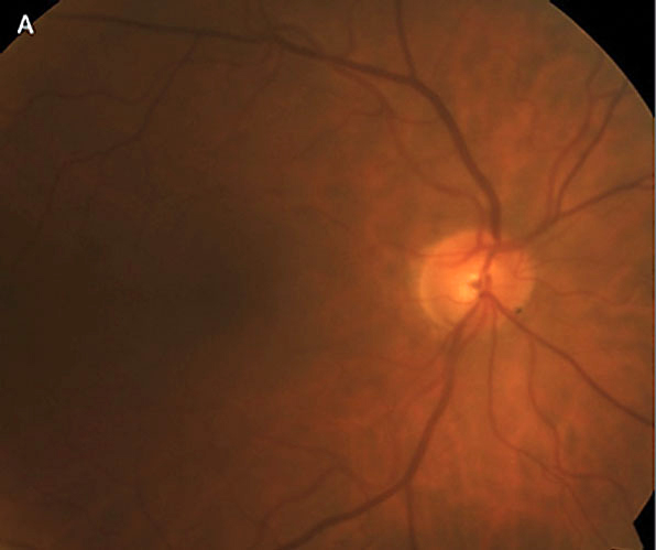
| Focal rim thinning (seen here at 7 o’clock) was associated with female sex and higher pattern standard deviation scores. Photo: Brian D. Fisher, OD. Click image to enlarge. |
The appearance of the optic disc is one of the key clinical signs used for diagnosing primary open-angle glaucoma (POAG). Disc appearance has been categorized into as many as four phenotypes in the past, most notably by Stephen Drance, namesake of the peripapillary RNFL hemorrhages he first identified. Each of these four phenotypes have been associated with certain systemic and ocular conditions as well as demographic factors, visual field patterns and disease prognosis. However, additional characteristic appearances have been described in the literature, including a pit-like area of ectasia in the lamina cribrosa. A reckoning is needed, experts now say.
To identify and better characterize various optic disc phenotypes in open-angle glaucoma, researchers examined 885 POAG eyes in a retrospective interventional case series. Based on patients’ disc photos, the researchers identified six distinct phenotypes and their distributions, according to their paper published last week in Eye. Phenotype characteristics and their prevalences were as follows:
- concentric rim thinning (18%), associated with thicker RNFL, higher mean deviation and lower pattern standard deviation compared with broad thinning
- focal rim thinning (45%), associated with female sex and higher visual field median pattern standard deviation
- acquired pit of the optic nerve (3%)
- tilted disc (12%), was associated with myopia, Asian race and younger age
- extensive peripapillary atrophy (5%), associated with older age
- broad rim thinning (17%).
“In an era of emerging technologies and increased access to a variety of devices to aid our diagnostic ability, we believe medical semiology remains an important tool for glaucoma specialists,” the researchers concluded in their paper.
Grassi L, Vega DS, De Gainza A, et al. Phenotypic expressions of the optic disc in primary open-angle glaucoma. Eye (Lond.) June 24, 2023. [Epub ahead of print]. |
