 |
|
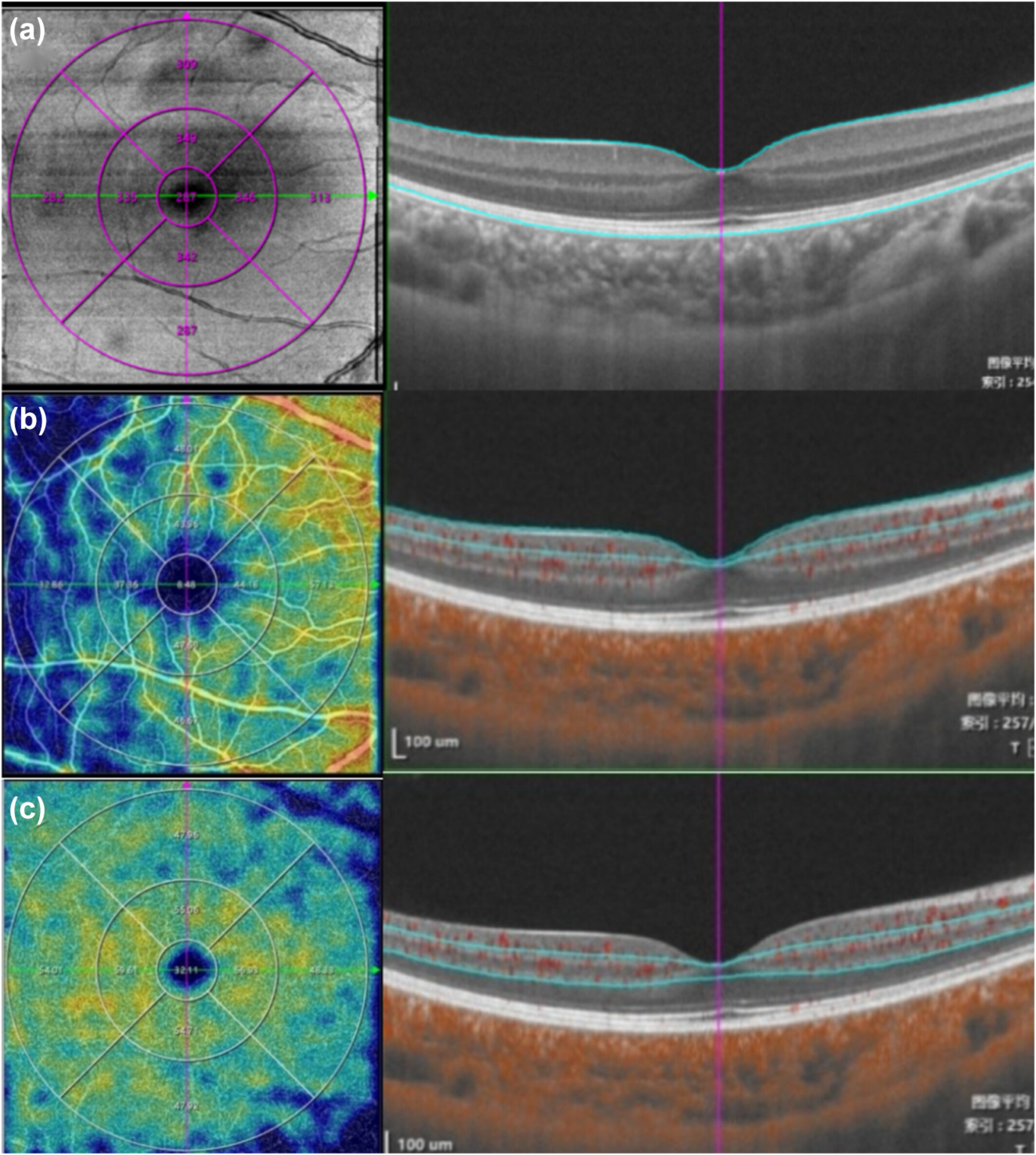
Pediatric axial myopic progression was associated with a decrease in choroidal thickness and retinal thickness in the parafoveal and perifoveal regions, while retinal thickness remained unchanged in the foveal area. This image from the study shows the segmentation of (a) total retinal thickness (b), superficial retinal vascular density and (c) deep retinal vascular density. Photo: Liu Z, et al. Ophthalmic Physiol Opt. February 7, 2024. Click image to enlarge. |
Since myopia typically develops in childhood, assessing the impact of myopia on retinal and choroidal structures and vasculature in myopic children is important. In a new study, researchers evaluated these structural features regions and their correlations with ocular biometric and vascular parameters in Chinese children using OCT angiography (OCT-A) and found that the thickness of retina and choroid was associated with axial length (AL), and strongly attributed to dynamic properties such as blood perfusion of the retina and choroid.
A total of 159 children between six and 13 years of age were included: 55 emmetropes (spherical equivalent refraction between -0.50D and +0.75D), 53 low-moderate myopes (between -0.50D and -6.00D) and 51 high myopes without pathological changes (myopia above -6.00D). Optical biometry was used to measure AL and anterior segment parameters. Swept-source OCT/OCT-A was used to assess the macular structures and vascular characteristics in a 6Å~ 6mm region centered on the macula.
This study showed that the thickness of the retina and choroid were not only associated with AL, but also showed dynamic properties such as the blood perfusion of the retina and choroid, particularly in the foveal area.
“The positive correlation between AL and macular blood perfusion is contrary to previously published studies on adults, which showed a negative association,” the authors explained in their article for Ophthalmic and Physiologic Optics. “One plausible explanation for this discrepancy may be due to the potential variability in the elasticity of fundus vessels between pediatric and adult subjects. This difference might contribute to variations in the autoregulatory capacity of these vessels, subsequently leading to metabolic changes in the retina."
Retinal thickness in the foveal area was positively associated with deep and superficial retinal vascular density, but not with AL. Deep retinal vascular density showed a more significant correlation than superficial retinal vascular density.
“Importantly, this relationship was independent of AL, as the previously observed positive correlation between foveal retinal thickness and AL disappeared after adjusting for the aforementioned factors,” the authors noted. “These results suggest that changes in blood perfusion might be the primary factor influencing alterations in RT in the central foveal area, for which there might be a compensatory increase in macular vascularization, ensuring adequate blood perfusion of the fovea to counteract the effects of excessive axial elongation. This mechanism likely plays a vital role in preserving the structural and functional integrity of the central region during the visual development of children with myopia.”
Boys exhibited significantly thicker RT in the foveal, parafoveal and perifoveal areas than girls, which is consistent with previous studies showing sex-related differences in macular thickness, with males having thicker retinas than females, the authors noted.
Liu Z, Liu M, Gou H, et al. Retinal and choroidal structure and vascularity in Chinese emmetropic and myopic children. Ophthalmic Physiol Opt. February 7, 2024. [Epub ahead of print.] |
