 |
|
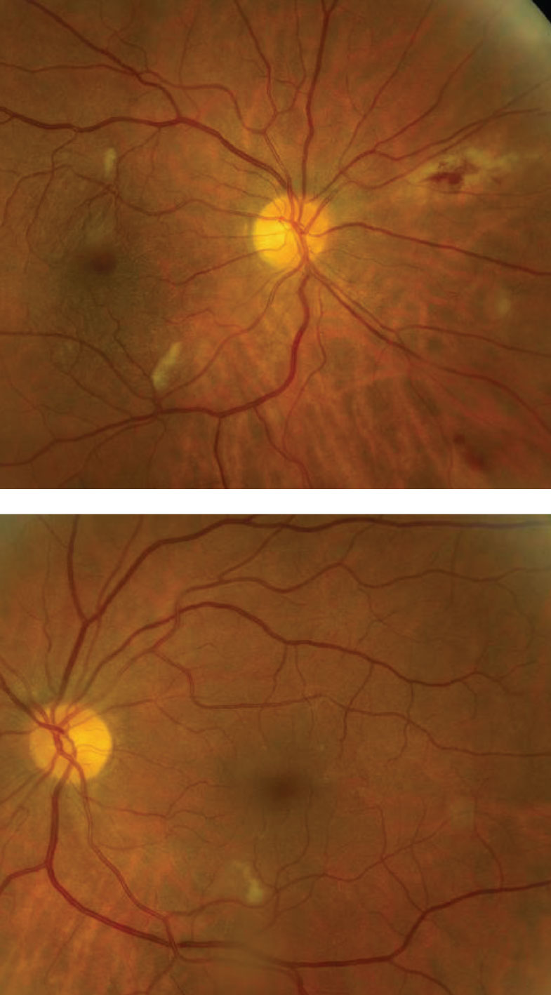
The cotton wool spots and other retinal findings observed in COVID-19 patients may actually be due to underlying conditions, research shows. Photo: Mark Dunbar, OD. Click image to enlarge. |
A recent study aiming to clarify and better understand the potential effect COVID-19 has on the retina did not find any retinal findings directly associated with the virus. Retinal findings that were noted in patients with COVID-19 were brought on by pre-existing conditions, the team noted.
A total of 119 eyes of 61 patients with a history of COVID-19 underwent color fundus photography and/or OCT of the macula within 90 days of their first positive test. The researchers assessed the presence of retinal heme, cotton wool spots, vascular sheathing and disc edema, as well as hyper-reflective changes, intraretinal fluid and subretinal fluid on OCT.
“Importantly, in a subset of eyes with no pre-existing conditions (many of which were contralateral ‘healthy’ eyes in patients with monocular conditions such as rhegmatogenous retinal detachment or retinal vein occlusion), no findings that could be associated with COVID-19 were seen,” the study authors concluded.
Previous studies have suggested retinal findings could be associated with COVID-19; however, many have questioned this hypothesis, raising the possibility that these changes may be attributed to retinal blood vessels.
“While similar appearing hyper-reflective structures were seen in this review of OCT imaging, all could be attributed to retinal blood vessels when correlated to simultaneous near infrared reflectance registration images of the retina,” the investigators explained. “This finding further calls into question the retinal manifestations of COVID-19 that were proposed early in the pandemic.”
Previous studies also examined patients with severe COVID-19, many of whom were in the ICU, and reported microvascular disease such as retinal hemorrhages and cotton wool spot, but this study wasn’t able to confirm these outcomes.
“It remains unclear if these microvascular changes can be attributed directly to COVID-19 or if they are a consequence of the generalized state of hemodynamic instability and heightened inflammatory response seen in many patients who require care in the ICU or are on positive pressure ventilation,” the authors noted.
Patel NS, Moon JY, Katz R, et al. Retrospective analysis of retinal imaging in COVID-19 positive patients at a tertiary eye care center. Clin Ophthalmol. 2021;15:3727-31. |
