 |
|
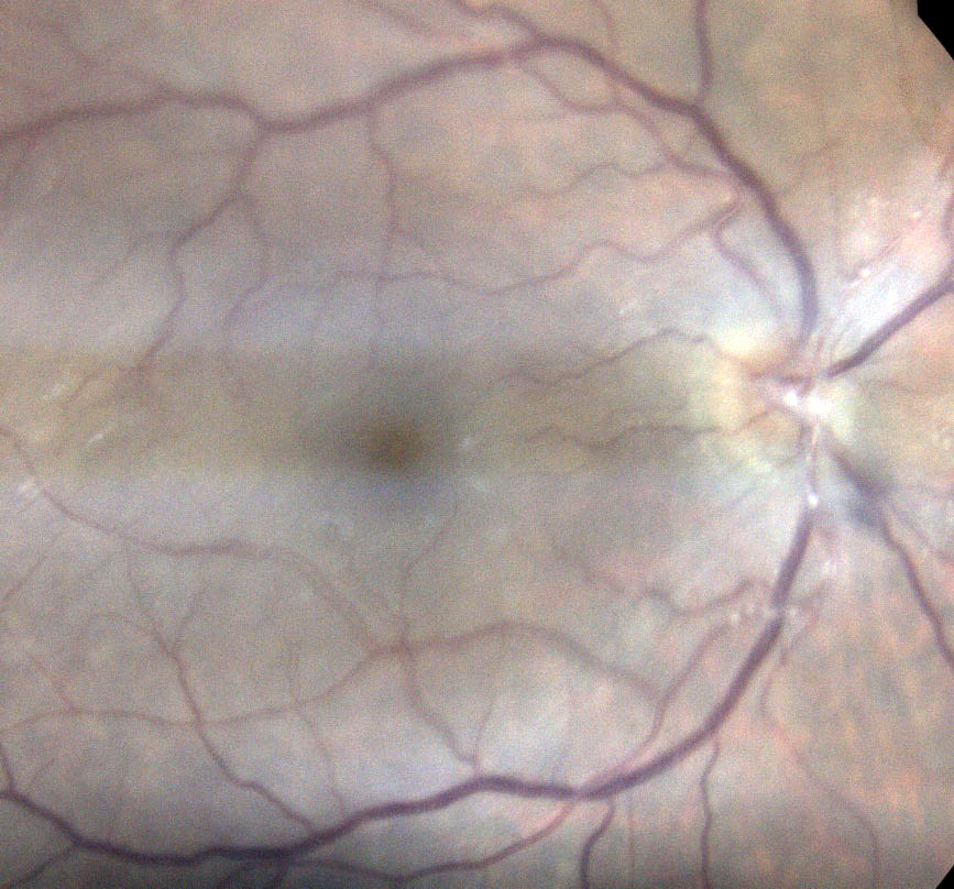
Occlusive arteritis of the primary retinal arterioles in a case of HSV keratouveitis. Photo: Aaron Bronner, OD. Click image to enlarge. |
Inflammation can be difficult to quantify in patients with uveitis, but interestingly, a new study presents evidence to suggest that it may correlate to retinal vessel diameter during active disease.
The findings showed that “the diameter of both retinal arteries and veins was increased in inflamed eyes, and this dilation reduced when the eyes were quiet,” the study authors reported in their paper. “These results suggest a possible correlation between retinal vessel diameters and the levels of intraocular inflammation.”
Color fundus images and clinical data of 89 eyes with uveitis were obtained from 70 patients across two visits, the first during the active disease stage (T0) and the second during inactive disease (T1). The researchers analyzed the change in central retinal vein equivalent (CRVE) and central retinal artery equivalent (CRAE) values from T0 to T1, taking into account clinical data such as age, gender, ethnicity, uveitis etiology and visual acuity.
From T0 to T1, the team observed a reduction in both CRVE and CRAE, “with active inflammation being able to influence the CRVE and CRAE after accounting for all other variables,” the authors noted. They determined that time was the only variable able to influence the degree of venular and arteriolar dilation, and BCVA was only influenced by time and ethnicity. The type of uveitis also had no influence on the observed correlation between retinal vessel diameter and disease activity.
The researchers explain in their paper that these findings demonstrate that systemic inflammation has a clinically relevant effect on the size of retinal vessels. “The results of the current study strengthen the hypothesis that retinal vessel diameters vary in response to inflammation and suggest that this could also be true if the inflammation is confined within the eye,” they explain.
Retinal vessel analysis may also assist in the objective grading of intraocular inflammation in patients with uveitis, “especially in challenging patients such as those with subclinical reactivations or showing lower degrees of inflammation,” the team concluded.
Zicarelli F, Agarwal A, Rizzi C, et al. Correlation between retinal vessel diameters and uveitis activity. Invest Ophthalmol Vis Sci. 2023;64(3):13. |
