 |
|
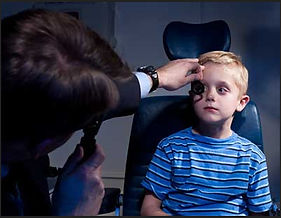
The interchangeability of retinoscopy and auto refractometry enhances the practical options available for measuring RPR in myopic patients in everyday practice. Photo: Longmeadow Optical. Click image to enlarge. |
Dust off your retinoscope if you want to have another tool available for assessing pediatric patients in a myopia management regimen. The increased emphasis on myopia of late has created a new slate of clinical responsibilities for many practitioners. With the concept of peripheral hyperopic defocus modification now part and parcel of myopia management strategies, clinicians need effective ways to measure relative peripheral refraction (RPR).
In a new study, researchers investigated whether retinoscopy provides RPR results comparable to autorefractometry in myopic individuals. Results showed that RPR is indeed a useful tool and may be used interchangeably with an autorefractor in everyday clinical practice.
Peripheral refraction was measured in 20 myopic kids and 20 controls. Both central and peripheral refraction were measured using retinoscopy and autorefractometry. Differences in the median central spherical equivalent, median RPR and median J45/J180 power vectors between the retinoscopy and autorefractometry techniques were analyzed.
In the myopic group, a typical small hyperopic shift in both the nasal and temporal peripheral retina for both methods were observed. Positive median values of RPR found was more than +0.50D and less than +1.0D, which is in agreement with previous studies on peripheral defocus in myopic subjects.
When RPR for the control group was analyzed, a small myopic defocus in the nasal and temporal retinal eccentricities was found, especially when using retinoscopy. “To our surprise, a small hyperopic RPR (median: +0.25D) in the control group was observed only in the nasal retina when autorefractometry method was used, but these values are within the measurement error range of the device,” the researched explained in their paper on the study. “Furthermore, we cannot exclude the effect of decreased repeatability of peripheral autorefraction measurements with increasing eccentricity. In general, the results related to RPR in emmetropic or hyperopic eyes are in a good agreement with other studies.”
Since not all myopes manifest peripheral hyperopic defocus, the ability to measure RPR in everyday practice may provide clinically relevant information of use in customizing optical interventions like orthokeratology, multifocal contact lenses and DIMS spectacles, all of which reduce the hyperopic defocus on the peripheral retina. Not all types of myopia show this characteristic of the retinal image in the eye, though. Retinoscopy allows for a quick assessment of the degree of peripheral defocus and make an informed decision as to whether these optical methods are indicated for the patient or whether pharmacologic therapy should be considered, the authors explained.
“Although retinoscopy requires patience and is a challenging task, our findings provide further evidence that it can be used interchangeably with the open-field autorefractometry technique when there is a need to examine peripheral refraction or RPR in myopic subjects,” the authors concluded. “This interchangeability of retinoscopy and open-field autorefractometry enhances the practical options available for measuring RPR in myopic subjects in everyday optometric or ophthalmic practice.”
The authors added that retinoscopy has several advantages over autorefractometry, such as cost-effectiveness, wider availability, mobility and more natural viewing conditions during testing.
Perdziak M, Prymula K, Krawczyk-Przekoracka A. Utility of retinoscopy to examine peripheral refraction. J Optom. December 30, 2023. [Epub ahead of print.] |

