 |
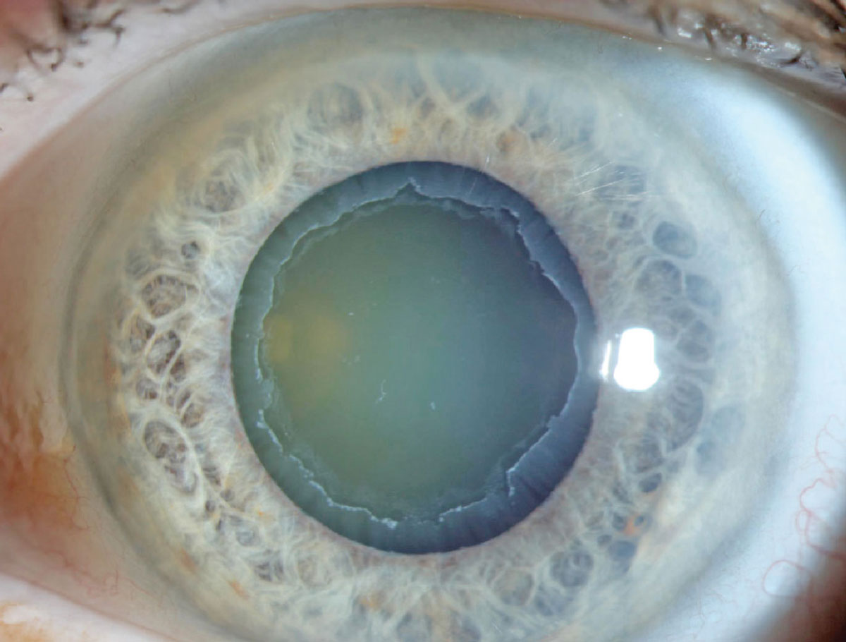
| OCT guided progression analysis showed that RNFL thinning progression appeared mainly in the superotemporal and inferotemporal areas in POAG and the temporal area in PXG. In both diseases, progressive GCIPL thinning was most common in the inferotemporal area. Photo: Aaron Bronner, OD. Click image to enlarge. |
Current research shows that more than half of patients with pseudoexfoliation syndrome—a systemic age-related disorder causing deposition of exfoliation material in ocular tissues and other organs—will go on to develop secondary open-angle glaucoma. Those who do will require frequent and diligent monitoring, as pseudoexfoliation glaucoma (PXG) is characterized by higher mean IOP levels, greater diurnal fluctuations in IOP and more marked pressure spikes than POAG. The condition has also been shown to result in progressively greater glaucomatous damage than POAG.
A recent study compared patterns of progressive RNFL and macular ganglion cell-inner plexiform layer (GCIPL) thinning in PXG vs. POAG patients using guided progression analysis on OCT (Cirrus HD). A total of 204 patients (204 eyes) were included in the study, of which 156 had POAG and 48 had PXG. The researchers observed several differences in progression patterns of the two groups, including more progressive RNFL and GCIPL thinning in PXG patients than in those with POAG.
Another difference involved the location of progressive thinning. “According to the RNFL progression-frequency maps, progression appeared mainly in the superotemporal and inferotemporal areas in POAG, whereas it had invaded more into the temporal area in PXG,” the study authors pointed out in their paper for Journal of Glaucoma. “According to the GCIPL maps, progression was most common in the inferotemporal area in both POAG and PXG.” They added that, when analyzing superior and inferior GCIPL thinning individually, the average progression rate was still faster in PXG than in POAG.
The analysis also showed that the range of long-term IOP fluctuation was greater in PXG, even though no significant difference was observed between average IOPs of the two groups during the follow-up period. The researchers argued in their paper that this factor may be the cause of the more progressive RNFL thinning seen in PXG patients, suggesting that “attention and effort are required not only to lower average IOP but also to reduce IOP fluctuation.”
In the future, studies with larger cohorts stratified by glaucoma stage would be beneficial to compare and better understand specific patterns of progressive RNFL and GCIPL thinning, the authors conclude.
Wy S, Lee YJ, Sun S, et al. Comparison of patterns of structural progression in primary open-angle glaucoma and pseudoexfoliation glaucoma. J Glaucoma. December 21, 2023. [Epub ahead of print]. |
