 |
|
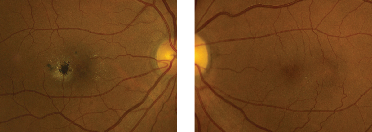
MacTel patients with the least severe disease demonstrated only key diagnostic features, while those with the most severe disease had both pigment and exudative neovascularization. Photo: Eric Dillinger, OD. Click image to enlarge. |
Affecting an oval-shaped area in the center of the fovea, macular telangiectasia type 2 (MacTel) is a rare retinal neurodegenerative disease that can cause vascular changes and lead to loss of visual acuity (VA). To improve the clinical understanding of MacTel, a team known as the MacTel Research Group has been studying the disease since 2005. With the goal of helping classify the disease and identify risk factors, the group recently performed an international multi-centered natural history study using spectral-domain OCT (SD-OCT). From the data, they were able to develop a scale to help characterize the severity of MacTel based on the amount of VA loss, ranging from least (grade zero) to most severe (grade six).
The study enrolled a total of 1,733 patients with MacTel from seven countries and 22 clinical sites. Multimodal imaging was performed on each patient, including stereoscopic color and red-free fundus photography, fluorescein angiography, fundus autofluorescence and SD-OCT. The images were subsequently analyzed using a machine learning algorithm.
The analyses validated three important features that may help classify disease severity and predict VA loss: OCT hyperreflectivity, pigment and ellipsoid zone (EZ) loss. The study authors noted that “by combining these three features (as absent, present, non-central involvement and central involvement of the macula), a seven-step scale was created, ranging from excellent to poor VA.” They explained that at grade zero, patients demonstrate only the key diagnostic features of MacTel (i.e., fluorescein angiographic leakage, retinal opacification, blunted or right-angle veins and blue-light reflectance); however, these patients lacked the three features noted above that were found to be significant for progression to VA loss. On the opposite end of the scale at the most severe grade (grade six), pigment and exudative neovascularization are present, which researchers explain is unsurprising given that those two events are known to cause significant vision loss.
In their paper, published in Ophthalmology Science, the researchers further described the clinical significance of the various MacTel classification grades that fall between zero and six. “In the presence of a non-foveolar EZ break (grade one), it is not surprising that the VA is unaffected; this begins in a noncentral location. However, when the EZ break affects the center of the fovea, the VA scores drop by almost 10 letters in grade two and 15 letters on average in grade three,” the team explained. “The next big step for VA decline is heralded by the presence of the OCT hyperreflectivity (grade four), followed by the presence of central pigmentary changes (grade five).”
By observing the presence or absence of hyperreflectivity, pigment and EZ loss on SD-OCT and referencing this newly established classification system, clinicians and researchers may be able to more effectively characterize the severity of MacTel and identify patients at risk of progression and VA loss.
Chew EY, Peto T, Clemons TE, et al. Macular telangiectasia type 2: a classification system using multi-modal imaging MacTel project report number 10. Ophthalmology. December 8, 2022. [Epub ahead of print]. |
