 |
| Decreased inferior RNFL thickness usually signifies progression, but it could also be an artifact of a poorly done scan. Photo: Danica J. Marrelli, OD. Click image to enlarge. |
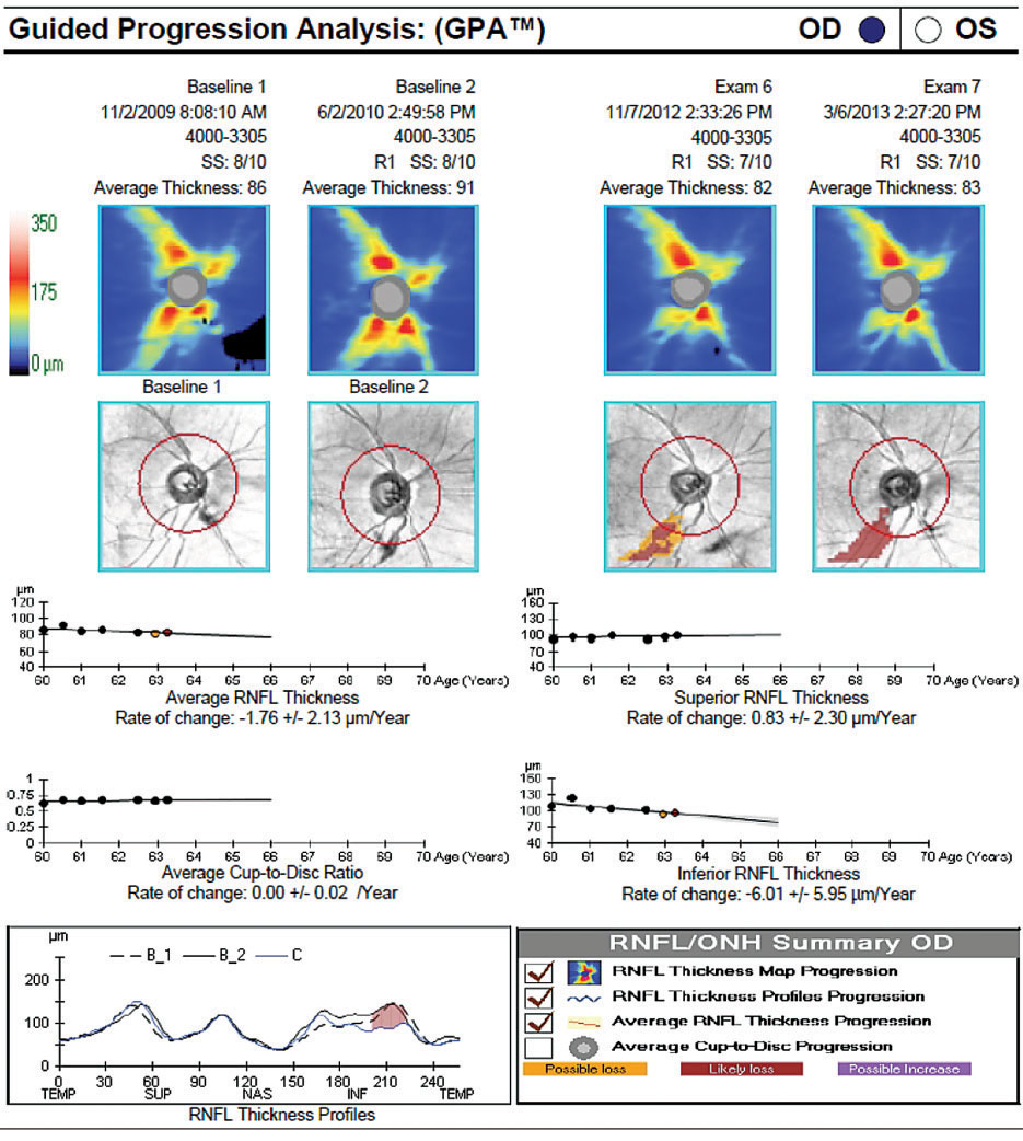
While it’s fair to say that the development of OCT radically improved diagnosis of glaucoma and assessment of its progression, the technology can sometimes be inconsistent in determining the appropriate scan location. Recognizing the need to better understand this problem, researchers recently initiated a retrospective, observational study to assess OCT guided progression analysis (GPA) in conditions with inconsistent scan locations.
The study included 84 glaucomatous eyes with at least four serial OCT tests. The researchers performed an analysis without manipulation—the control set—and then again after the OCT scan of the latest test was intentionally moved inferotemporally—the test set. Their goal was to evaluate the ability of GPA to adjust the OCT scan location. They classified the eyes into adjustment and non-adjustment groups based on the superior or inferior quadrant retinal nerve fiber layer (RNFL) thickness agreement between the two sets.
The researchers compared GPA parameters between the sets and found that the test set showed a bigger superior RNFL thickness and smaller inferior RNFL thickness vs. the control set. Additionally, the test set showed a greater rim area, average cup-to-disc ratio and cup volume than the control set.
GPA automatically adjusts the scan location in agreement with the previous location using one of two registration methods, the study authors explained. “R1” is registration based only on the translation of the disc center, and “R2” is registration based on the translation and rotation of the OCT fundus.
The researchers observed that the eyes in the non-adjustment group had a lower chance of applying the eye-tracking function and a higher frequency of the “R1” registration method of OCT GPA than the adjustment group. They also reported that all eyes with the “R1” method were in the non-adjustment group while all eyes with the “R2” method were in the adjustment group.
“Changes in the OCT scan location induced changes in RNFL thickness that were not completely compensated for, despite the use of OCT GPA, especially when the “R1” registration method was used or the eye-tracking function was not available during the image acquisition,” the study authors concluded. “On the contrary, the “R2” method showed a successful adjustment of the scan location in all cases. Regardless of the registration method, the optic nerve head parameters were not adjusted. These findings may lead to the misidentification of the detection of progression.”
Hwang YH, Song MY. Effect of inconsistent optical coherence tomography scan location on glaucoma progression analysis. J Glaucoma. February 18, 2022. [Epub ahead of print]. |
