 |
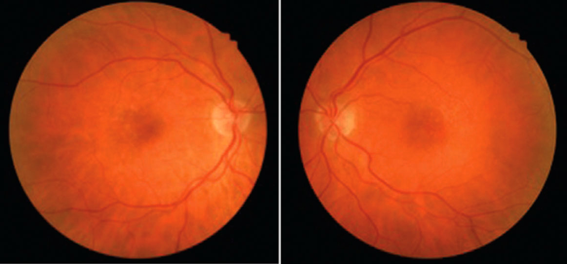
| This study identified various biomarkers of progression from early to intermediate AMD. Photo: Amanda Legge, OD. Click image to enlarge. |
Therapeutic development in dry age-related macular degeneration (AMD) has proven difficult due to the lack of available biomarkers to help monitor progression of disease in its early stages. To address this, researchers initiated a study evaluating visual function changes in early and intermediate AMD. They found that AMD progression is slow and appeared not to be linear over the 24-month study period.
This prospective, observational study recruited 101 subjects, including 33 early AMD, 47 intermediate AMD and 21 healthy participants. Longitudinal changes in visual function metrics over 24 months was the primary objective. The visual function tests conducted included best-corrected visual acuity, low luminance visual acuity, microperimetry, cone contrast testing and dark adaptation. The team also tested whether a limited number of genetic risk alleles as well as reticular pseudodrusen (RPD) and hyperreflective foci (HRF) were predictors of diagnosis and visual function performance.
Of the recruited participants, 70 completed the two-year visit (22 early AMD, 31 intermediate AMD and 17 controls). The researchers found that the percent reduced threshold on microperimetry and cone contrast testing red significantly distinguished intermediate AMD when compared with controls after 12 and 24 months, respectively.
The analysis also demonstrated that cone contrast testing red, percent reduced threshold and absolute threshold on microperimetry showed a significant longitudinal deterioration of visual function in intermediate AMD vs. controls at 12 months and beyond. However, the researchers noted, there was a reduced rate of worsening.
The findings also showed that dark adaptation data confirmed a pre-existing functional deficit among intermediate AMD patients at baseline. Additionally, the study authors observed that HRF and RPD are moderately linked with visual function decline measured by dark adaptation.
“In intermediate AMD, microperimetry variables, cone contrast testing red and dark adaptation revealed slow and non-linear functional decline over 24 months. A structure-function relationship in early-intermediate AMD stages was demonstrated between HRF, RPD and dark adaptation, possibly modified by genetic risk factors,” the study authors concluded. “These structural and functional features represent potential endpoints for clinical trials in intermediate AMD.”
Lad EM, Fang V, Tessier M, et al. Longitudinal evaluation of visual function impairments in early and intermediate age-related macular degeneration patients. Ophthalmology. May 20, 2022. [Epub ahead of print]. |

