No one would dispute that dilation is indicated for a patient who presents with acute-onset photopsia in the presence of floaters—but would you dilate a healthy, asymptomatic 25-year-old female optometrist with best-corrected visual acuity of 20/15 in each eye who presents solely for an updated glasses prescription?1 Would your recommendation change if she had refractive error of -7.00D in each eye, and her last normal peripheral retinal examination was performed one year prior?
Advances in technology have given optometrists the ability to better view the posterior segment without subjecting patients to dilation. In particular, the development of ultra-widefield imaging (UWFI) by companies such as Optos, Centervue and Heidelberg Engineering provides optometrists with an adjunctive tool of objective retinal documentation. For asymptomatic patients undergoing routine eye examination, we may ask ourselves if dilation is really necessary. Are ancillary procedures such as UWFI appropriate substitutes for dilation in low-risk patients?
Pupillary dilation is essential for the thorough stereoscopic assessment of ocular health, including peripheral fundus examination.2 In the state of Florida, pupillary dilation is required by law for all patients undergoing an initial comprehensive eye examination.3
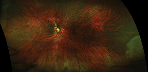 |
| Ultra-widefield image of clinically diagnosed inferior retinal break with shallow retinal detachment not apparent in image. Click image to enlarge. |
Given that malpractice litigation against optometrists is frequently based on the misdiagnosis of retinal pathology due to failure to dilate, and that eye care professionals have had access to diagnostic topical agents in some states since the early 1970s, it is striking that such significant dialogue in eye care still exists regarding the pertinence and frequency of routine dilation.4,5
How Frequent is Enough?
Evidence-based clinical practice guidelines on the frequency of dilated fundus examinations have been well established in patients with concomitant systemic disease (e.g., diabetes mellitus) and ocular disease risk factors such as primary open angle glaucoma, lattice degeneration and posterior vitreous detachment.1,6
The American Optometric Association’s 2015 evidence-based clinical practice guideline states that pharmacological dilation is generally required for the thorough evaluation of ocular structures.2 For patients between the ages of 18 and 39, a comprehensive eye examination including ocular health evaluation is recommended at least every two years.2 For patients age 65 and older, comprehensive eye examinations are recommended annually in the absence of a diagnosed ocular condition.2 More frequent monitoring with dilation is indicated in a patient with a previous diagnosis of ocular pathology, or if the patient is at risk for developing ocular disease, and if there is a change in patient symptomatology such as new onset photopsia, floaters or vision loss1,2,7,8
Although the presence of symptoms—including visual changes, flashes of light or floaters—may affect the frequency of dilation, it should not be the only indication for dilation of a patient otherwise considered to be at “low risk” for retinal pathology.1,2
In a population-based analysis of patients age 40 or older, 2.39% of patients with normal baseline eye examination experienced vision loss (visual acuity less than 20/40 or visual field loss) over a five-year period without follow-up examination that included dilation during that time period.7 Researchers recently determined that only 4% of adult patients who presented for routine eye exam had peripheral retinal pathology requiring treatment that would have gone undiagnosed without pupillary dilation.8 As such, the clinical utility and cost effectiveness of routine dilation, in the absence of a known pathology, is sometimes brought into question.7,8 It is increasingly common for some practitioners to offer UWFI as an alternative to a dilated exam. Doing so must include a full explanation of the trade-offs, and this does not absolve you of responsibility to arrive at a diagnosis even in the absence of evidence of pathology on UWFI.
Keep in mind that, although helpful, guidelines are just that. Clinical acumen will guide the practitioner when more frequent dilation is necessary. However, if in doubt, the most appropriate course of action is to dilate for all of your fundus examinations.
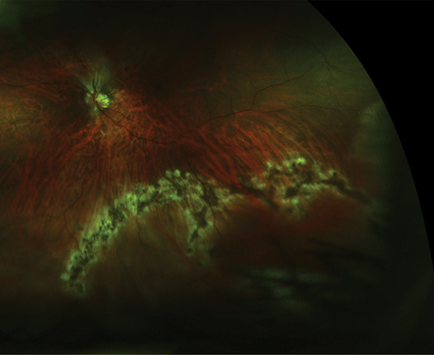 |
| Post-treatment UWFI of the same patient. Click image to enlarge. |
UWFI vs. Peripheral Exam?
Ultra-widefield imaging devices have the capacity to document peripheral retinal pathology, providing up to a 200-degree temporal and nasal imaging field and the ability to image up to 82% of the retina.9 However, retinal lesions located anterior to the equator are likely to be missed by doctors using UWFI alone.10 Researchers have determined this technology to have reduced sensitivity (36%) for the detection of the lesions compared with binocular indirect ophthalmoscopy using scleral indentation (76%), and concluded that ultra-widefield imaging alone is not sufficient for making a clinical diagnosis.10
Advantages of UFWI
Applications of ultra-widefield imaging have been expanded and applied to fluorescein angiography (FA), indocyanine green angiography, and fundus autofluorescence (FAF).9 In patients with diabetes, UWFI in fluorescein angiography has been proven to be advantageous over the seven standard mydriatic stereoscopic 30-degree images that were used by the Early Treatment Diabetic Retinopathy Study (ETDRS).11,12 Research comparing ultra-widefield and standard techniques identified retinal non-perfusion and neovascularization in an additional 10% of eyes, which would have otherwise been missed with standard imaging techniques alone.11
Research also shows that non-mydriatic ultra-widefield imaging correlates well with the ETDRS seven standard 30-degree field fundus photography for the identification of diabetic retinopathy.13 Additionally, diabetic retinopathy was identified 17% more frequently in non-mydriatric ultra-widefield imaging as compared with the ETDRS standard.14,15
Limitations of UWFI
UWFI possesses its own set of drawbacks relative to traditional fundus photography methods. The lack of “true-color” images, due to the use of red and green scanning lasers, may limit interpretation compared with traditional fundus photography.9
The technology’s most clinically significant limitation is its inability to image the entire retina.9 Recall that 18% of the retinal area cannot be imaged through a dilated pupil with current ultra-widefield technology.9 Additionally, the lack of sensitivity to retinal lesions anterior to the equator compared with clinical examination means that UWFI is not apt to detect retinal holes and breaks, especially anterior to the equator in the superior and inferior quadrants, and is therefore recommended only as an adjunct to a dilated fundus exam that includes careful peripheral retinal examination.9,10,16
The Role of Ancillary Testing
To patients and professionals alike, new technology is often equated with better quality of care.17 However, the cost of new devices may create financial pressure for practitioners to incorporate their use into routine evaluation.18 Performing ancillary tests for screening purposes without regard to a patient’s individual clinical presentation or clinical history without reasonably expected benefit can be construed as unnecessary and unethical.19 This may be particularly concerning when practitioners profit from performing such tests.18-20
Ancillary procedures that lack a medical indication are not covered by most insurance plans, including Medicare, requiring the overall cost to be borne by the patient. If done under circumstances that do not offer a patient benefit, such testing runs the risk of been called exploitative.18-20 Of course, on an individual basis, the same test may indeed be necessary or justifiable. For instance, UWFI screening may reveal pathology or raise suspicion that warrants additional clinical investigation, or the patient may prefer the convenience of avoiding a dilated exam and be willing to accept it when properly educated about the different testing methods available. Regardless, the determination of necessity of ancillary testing cannot occur without a complete clinical evaluation.20,21
The standard of care in medical practice evolves from the behavior of physicians.22 Choosing to perform ancillary tests for screening purposes should not supplant a thorough clinical exam and full patient history; otherwise, the result could influence one’s diagnosis or management and can establish incomplete medical decision-making protocols.19,20,22
Case Outcome
Our optometry colleague-turned-patient agreed with her OD’s recommendation for routine dilation. Stereoscopic evaluation of the far inferior periphery of the left eye revealed pinpoint vitreous hemorrhage with associated retinal tear, and shallow retinal detachment. Documentation of the lesion with UFWI was unsuccessful because the pathology was located outside the field of view. She was promptly treated with laser photocoagulation and maintained visual acuities of 20/15 in each eye.
In this case, not only was treatable pathology uncovered in a healthy, asymptomatic patient that would have gone undiagnosed without routine dilation, but the lesion was not visible with UWFI—a close-to-home example of the technology’s limitations as a primary diagnostic modality. Although cases of treatable, vision-threatening peripheral retinal pathology in asymptomatic patients are uncommon, this particular one—involving an OD—allows us to address the debate from a distinct perspective: If it were your eye, would you dilate? Therein lies the critical importance that education and consent play. Many low-risk patients do elect to choose UWFI as a time-saving alternative to a dilated exam without incident, after giving their informed consent, while more risk-averse patients may decline.
UWFI, including its application to fluorescein angiography and other diagnostic modalities, has provided optometrists with an expanded, objective way to document posterior segment findings as well as to better understand the role of peripheral pathology in retinal disease.9 UWFI should augment, not replace, your own clinical acumen. Like any other diagnostic test, it should be used as a tool—not a crutch.
Dr. Steen is an attending optometrist and instructor of ocular pharmacology at Nova Southeastern University’s College of Optometry.
|
1. American Academy of Ophthalmology preferred practice patterns committee. Preferred Practice Pattern Guidelines. Posterior vitreous detachment, retinal breaks and lattice degeneration. San Francisco, CA; American Academy of Ophthalmology; 2014. Available at www.aao.org/ppp. 2. AOA Evidence-based optometry guideline development group. Evidence-Based Clinical Practice Guideline. Comprehensive Adult Eye and Vision Examination. American Optometric Association. St. Louis, MO; 2015. Available at www.aoa.org. 3. Florida Department of State. Board of Optometry Standards of Practice. Chapter 64B13-3. Available at www.flrules.org/gateway/ruleno.asp?id=64B13-3.007. 4. Classé JG. A review of 50 malpractice claims. J Am Optom Assoc. 1989;60:694-706. 5. Classé JG. Liability and opthalmic drug use. Optom Clin. 1992;2(4):121-34. 6. American Academy of Ophthalmology preferred practice patterns committee. Preferred Practice Pattern Guidelines. Primary Open Angle Glaucoma. San Francisco, CA; American Academy of Ophthalmology; 2015. Available at www.aao.org/ppp. 7. Taylor HR, Vu HT, McCarty CA, Keeffe JE. The need for routine eye examinations. Invest Ophthalmol Vis Sci. 2004;45(8):2539-42. 8. Siegel BS, Thompson AK, Yolton DP, et al. A comparison of diagnostic outcomes with and without pupillary dilation. J Am Optom Assoc. 1990;61(1):25-34. 9. Shoughy S, Arevalo JF, Kozak I. Update on wide- and ultra-widefield retinal imaging. Indian J Ophthalmol. 2015;63(7):575-581. 10. Mackenzie PJ, Russell M, Ma PE, et al. Sensitivity and specificity of the Optos Optomap for detecting peripheral retinal lesions. Retina. 2007; 27(8):1119-24. 11. Wessel MM, Assker GD, Parlitsis G, et al. Ultra-wide-field angiography improves the detection and classification of diabetic retinopathy. Retina. 2012;32:785-91. 12.Tan CS, Sadda SR, Hariprasad SM. Ultra-widefield retinal imaging in the management of diabetic eye disease. Ophthalmic Surg. Lasers Imaging Retina. 2014;45:363-6. 13. Kernt M, Hadi I, Pinter F, et al. Assessment of diabetic retinopathy using nonmydriatic ultra-widefield scanning laser ophthalmoscopy (Optomap) compared with ETDRS 7-field stereo photography. Diabetes Care. 2012;35:2459-2463. 14. Silva PS, Cavallerano JD, Sun JK, et al. Nonmydriatic ultrawide field imaging compared with dilated standard 7-field 35mm photography and retinal specialist examination of diabetic retinopathy. Am J Ophthalmol. 2012;154:549-559. 15. Silva PS, Cavallerano JD, Tolls S et al. Potential efficiency benefits of nonmydriatic ultrawide field retinal imaging in an ocular telehealth diabetic retinopathy program. Diabetes Care. 2014;37:50-55. 16. Kornberg DL, Klufas MA, Yannuzzi NA, et al. Clinical utility of ultra-widefield imaging with the Optos Optomap compared with indirect ophthalmoscopy in the setting of non-traumatic rhegmatogenous retinal detachment. Semin Opthalmol. 2015; 21:1-8. 17. Deyo RA. Cascade effects of medical technology. Annu Rev Public Health. 2002;23:23-44. 18. An Optometrist’s Guide to Clinical Ethics. Bailey RN, Heitman E, eds. St. Louis, MO: American Optometric Association, 2000. 19. Advisory Opinion of the Code of Ethics. Appropriate examination and treatment procedures. San Francisco, CA: Am Acad Ophthal; 2007. Available at www.aao.org/ethics-detail/advisory-opinion--appropriate-examination-treatmen. 20. Augsburger JJ. Unnecessary clinical tests in ophthalmology. Trans Am Ophthalmol Soc. 2005;103:143-47. 21. Mold JW, Stein HF. The cascade effect in the clinical care of patients. N Engl J Med. 1986 20;314(8):512-4. 22. EL Raab. Peer discussion: Augsburger JJ. Unnecessary clinical tests in ophthalmology. Trans Am Ophthalmol Soc. 2005;103:143-47. |
