 |
Fibrovascular proliferation in the eye is a pathological process characterized by the growth of fragile new vessels together with fibrous scar tissue. While angiogenesis is a normal response to injury in many tissues, neovascularization with fibrovascular growth can lead to serious vision-threatening consequences, such as intraocular hemorrhage, inflammation, traction and secondary glaucoma. As such, it is important to understand where fibrovascular tissue arises in the eye, its histological components, how to identify it and the underlying causes that trigger its formation.
Anatomical Sites of Growth
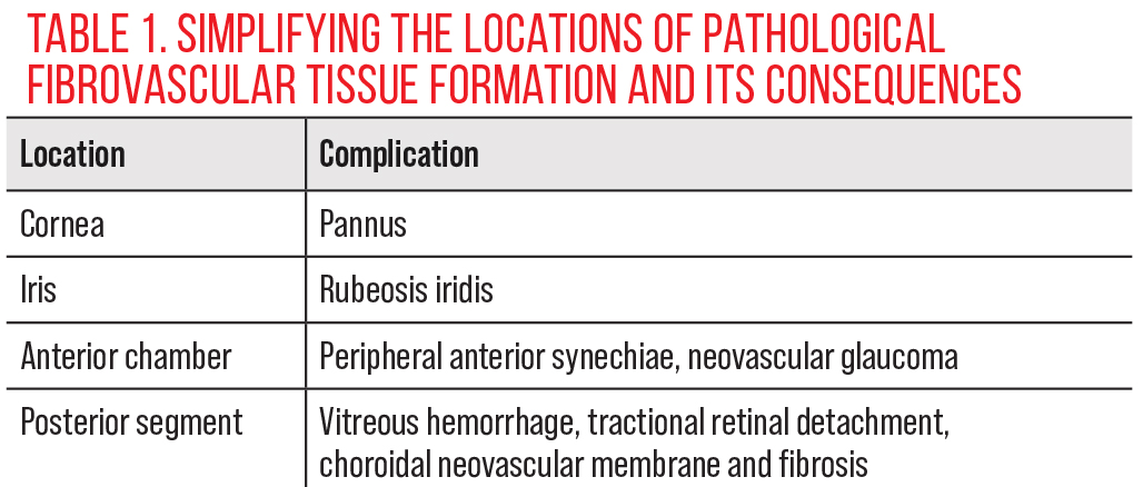
Fibrovascular membranes can develop in multiple regions of the eye and to variable degrees, depending on the underlying pathological stimulus and affected area.
• Cornea. Although the cornea is avascular by design, there are pathological conditions that initiate the growth of fibrovascular tissue. Commonly, this can include contact lens–induced hypoxia, chemical burns, bacterial or viral corneal infections and immune diseases such as Stevens-Johnson syndrome. These inciting conditions cause breakdown of the limbal barrier, permitting stromal invasion by blood vessels. As a result, fibroblasts deposit collagen and lead to the formation of pannus, which is defined by neovascularization with abnormal tissue. Ultimately, this threatens transparency of the cornea.1
• Rubeosis iridis. Iris neovascularization can also lead to fibrovascular tissue growth in the anterior segment. Ischemic diseases of the retina, such as diabetic retinopathy (DR) or ciliary body ischemia, can cause vascular endothelial growth factor (VEGF) to diffuse into the anterior segment of the eye, leading to capillary formation on the pupillary margin. Fibrous tissue forms around these vessels. Complications can include bleeds, such as in hyphema, as well as inflammation from vascular permeability. Continued growth can invade the angle.2
 |
|
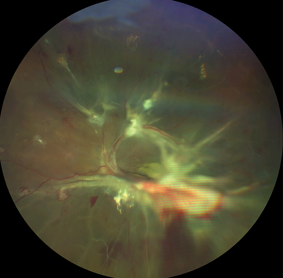
This fundus photo shows white fibrous tissue membranes accompanying neovascularization in proliferative DR. Click image to enlarge. |
• Anterior chamber angle. Abnormal growth of vessels can also invade the trabecular meshwork, accompanied by contractile fibrous tissue. It is these fibrovascular membranes that mature further and contract to form peripheral anterior synechiae, ultimately leading to angle closure and neovascular glaucoma.3
• Vitreous and retina. Fibrovascular tissue may develop on the surface of the retina or along the posterior hyaloid face of the vitreous. This is of particular importance in proliferative retinal disease (e.g., DR), where fibrovascular tissue accompanies neovascularization and can extend into the vitreous. These are often visually catastrophic, as the new blood vessels can bleed and contract, causing hemorrhage or retinal detachment.4-6
• Choroid /sub-RPE space. In some contexts, particularly in neovascular age-related macular degeneration, fibrovascular tissue can grow from the choroid through or under the retinal pigment epithelium (RPE), forming choroidal neovascularization and fibrosis.7
 |
|
Click table to enlarge. |
Histological and Cellular Components
Fibrovascular growth in the eye is typically initiated by hypoxia and tissue damage, which lead to an imbalance of pro- and anti-angiogenic factors. There are many pathological ocular conditions that trigger this process, varying by location.
Fibrovascular membranes are composed of a mixture of cellular and extracellular elements. This often includes abnormal growth of new blood vessels that are typically thin-walled, fragile, lacking a robust muscular layer and prone to breaking/leakage. Additional components include fibroblasts and myofibroblasts that function to produce extracellular matrix, contract and remodel tissue. This extracellular matrix consists of collagen and other proteins that provide structural support to the fibrovascular membrane. Finally, depending on the cause and chronicity, inflammatory cells may also be present. The relative proportion of vessels vs. fibrous tissue depends on the stage and etiology, where newly formed membranes may be more vascular and chronic ones often become fibrotic and contractile.1-3
Clinical Identification
Recognizing fibrovascular growth early is critical, as timely intervention can prevent vision loss. Depending on its location, there are many methods for evaluation.
For anterior segment conditions, slit lamp examination with gonioscopy is vital in determining its presence and the potential underlying condition. Neovascularization with accompanying fibrovascular growth on the iris appears as fine, abnormal blood vessels on the iris surface, often near the pupillary margin. Gonioscopy can visualize if these new vessels extend into the angle, sometimes with an overlying fibrovascular membrane. Over time, membranes may contract, causing synechiae that are also visible on gonioscopy.
In proliferative retinal diseases, neovascularization on the optic disc, elsewhere or on the retinal surface can be seen with indirect ophthalmoscopy, particularly after dilation. Fibrovascular membranes may appear as grayish-white elevated tissue, often with looping abnormal vessels, preretinal hemorrhages and tractional folds. Oftentimes, when neovascular vessels are fine and not easily observed, the fibrovascular tissue is more identifiable and suggestive of proliferative disease.
Using advanced imaging such as fluorescein angiography, dye leakage from abnormal vessels helps identify neovascular areas. OCT angiography provides high-resolution, noninvasive imaging of blood flow in the retina and vitreous, enabling detection of neovascularization and fibrovascular proliferation, even before they become clinically obvious. Structural OCT may show hyperreflective membranes on the retinal or vitreous surface.
Takeaways
Fibrovascular tissue growth in the eye is a pathologic process that arises in response to ischemia, inflammation, trauma or immune conditions. These membranes can form in the iris, angle, retina, vitreous or subretinal spaces, depending on the underlying process. Understanding the mechanisms and early detection of fibrovascular proliferation is critical because timely intervention can preserve vision and prevent irreversible damage.
Dr. Labib graduated from Pennsylvania College of Optometry and is now dean of Pacific University's College of Optometry. She completed her residency in primary care/ocular disease and is a fellow of the American Academy of Optometry and a diplomate in the Comprehensive Eye Care section. She has no financial interests to disclose.
1. Espana EM, Di Pascuale MA, He H, et al. Characterization of corneal pannus removed from patients with total limbal stem cell deficiency. Invest Ophthalmol Vis Sci. 2004;45(9):2961-6. 2. Pagoulatos D, Georgakopoulos C. Rubeosis iridis. Pan Afr Med J. 2017;28:279. 3. Senthil S, Dada T, Das T, et al. Neovascular glaucoma - a review. Indian J Ophthalmol. 2021;69(3):525-34. 4. Stewart JM, Coassin M, Schwartz DM. Diabetic retinopathy. In: Feingold KR, Ahmed SF, Anawalt B, et al., ed. Endotext [Internet]. South Dartmouth (MA): MDText.com. Updated May 14, 2025. www.ncbi.nlm.nih.gov/books/NBK278967/. Accessed December 5, 2025. 5. Elborgy E, Pulido JS. Posterior pole and peripheral retinal fibrovascular proliferation in von Hippel Lindau disease. Asia Pac J Ophthalmol (Phila). 2017;6(3):256-60. 6. Sawa H, Ikeda T, Matsumoto Y, et al. Neovascularization from scleral wound as cause of vitreous rebleeding after vitrectomy for proliferative diabetic retinopathy. Jpn J Ophthalmol. 2000;44(2):154-60. 7. Bhatt NS, Diamond JG, Jalali S, Das T. Choroidal neovascular membrane. Indian J Ophthalmol. 1998;46(2):67-80. |