 |
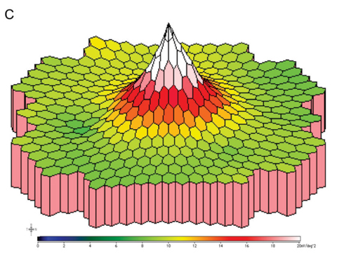
A reliable way to preoperatively assess the fundus and detect related disease in patients with turbid lenses is with full-field electroretinogram and multifocal electroretinogram (pictured: mfERG three-dimensional P1 response density plot). Photo: Hua Bi, OD, PhD. Click image to enlarge. |
For patients who wait until their cataracts are severe to seek treatment, it can become extremely challenging to evaluate the structure and function of the fundus due to the opacity of the lens. At the same time, this assessment is imperative to ensure a patient does not have fundus disease prior to undergoing surgery. Ocular B-ultrasound is one method to screen for fundus disease in eyes with a turbid lens; however, its accuracy in diagnosing retinopathy is not high. A potential alternative that cataract surgeons have been investigating in the last decade is objective visual electrophysiology technology, which involves a series of noninvasive tests to provide objective indicators of visual system functions. One recent study found that even in mature cataract patients, this tool was effective in evaluating fundus function and could be used to detect fundus disease.
A total of 124 mature cataract patients (153 eyes) completed pre-cataract surgery examinations, including BCVA, pattern visual evoked potential (PVEP), full-field electroretinogram (fERG) and multifocal electroretinogram (mfERG). Postoperatively, patients were divided into two groups based on whether fundus disease was detected. One month after surgery, BCVA was measured, and visual electrophysiology was performed on subjects who had a stable fundus condition and had not been treated for fundus disease.
Compared with the group without fundus disease, the researchers noted several differences in the preoperative electrophysiological exam of the group with fundus disease, such as that the amplitude of fERG waves and the amplitude density of the P1 wave in the 2nd to 5th rings of mfERG were decreased.
Visual electrophysiology was also able to predict postoperative vision in mature cataract patients. The following parameters were negatively correlated with postoperative BCVA:
- In the group without fundus disease: the amplitude of the PVEP 15′ P100 wave and the amplitude of dark-adapted (DA) 0.01 b-wave, DA 3.0 a-wave, and DA 10.0 a-wave
- In the group with fundus disease: the amplitude of PVEP and fERG and the amplitude density of mfERG
While the authors reported that based on the study’s findings, fERG and mfERG can be used for differential diagnosis of fundus disease, they noted that PVEP did not show significant diagnostic value.
The researchers also observed that different types of cataracts have different effects on electrophysiological examination results. In their paper, published in the Journal of Ophthalmology, they elaborated on this finding: “In the eyes of cortical cataracts, some parameters of PVEP, fERG, and mfERG were significantly different before and after surgery. In the eyes of nuclear cataracts, some parameters of fERG and mfERG were significantly different before and after surgery.” Finally, “in the eyes of posterior subcapsular cataracts, some parameters of PVEP and fERG were significantly different before and after surgery.”
Based on these findings, the study authors advise that both fERG and mfERG can be used to detect fundus disease in mature cataract patients. “The preoperative visual electrophysiological examination has high clinical value in predicting postoperative vision of mature cataract patients with fundus disease. When interpreting the electrophysiological report, it is necessary to consider the existence of cataracts,” they concluded.
Zhang M, Ji M, Tan M, Yu Y, Guan H. Evaluation of fundus function in mature cataract patients by visual electrophysiology. J Ophthalmol. October 31, 2023. [Epub ahead of print]. |
