 |
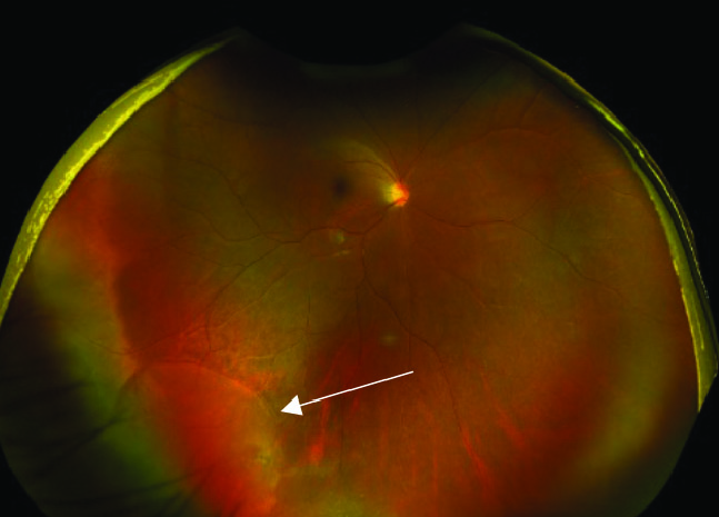
| UWF imaging is capable of detecting retinal tears and holes but some may be missed without acquiring additional photos at different angles, the study found. Photo: Brad Sutton, OD. Click image to enlarge. |
The usage of ultra-widefield imaging (UWF) is becoming more prevalent, despite concerns that it can miss retinal lesions that are located anterior to the equator. In a new study, researchers found that UWF showed encouraging results in detecting peripheral retinal tears and holes, though it did miss about one-third of such lesions.
A total of 198 patients diagnosed with acute posterior vitreous detachment were included. Eyes were divided into two groups: 89 with peripheral retinal holes and tears treated with laser retinopexy (treatment group) and 109 controls. Patients underwent UWF imaging and indirect ophthalmoscopy with scleral depression. UWF images from both groups were reviewed by two blinded graders and then compared to funduscopic exam and medical records.
UWF imaging showed moderate sensitivity (67.4%) and high specificity (98.2%) for detecting treatment requiring peripheral retinal lesions, with most misses occurring anterior to the equator and more frequently superiorly, inferiorly or nasally, the authors explained. The inter-rater agreement was high at 87.9% which is consistent with previous data.
The lesions anterior to the equator were the most likely to be missed by a grader on UWF imaging, showing the importance of scleral depressed exam as the diagnostic standard of care.
The authors point out multiple reasons why there was only moderate sensitivity of detecting treatment requiring retinal lesions. “For one, each Optomap image is large (2000x2000 pixels) and can show 200° view of the retina, with an angular resolution of 12 pixels per degree of retina. Accounting for the distance from the optic nerve to the fovea, this translates to approximately 200 pixels. This equates to a relatively low resolution for the detection of small peripheral retinal lesions,” the authors explained. The low pixel density also results in decreased contrast sensitivity when evaluating a small hole and adjacent normal retina. “The combined low resolution and the resultant decreased contrast sensitivity are likely contributing factors to the majority of retinal lesions that were missed posterior to the equator.”
For retinal lesions anterior to the equator, challenges of imaging lesion in the far periphery could be the attributed to patient positioning, inferior eyelash artifact or media opacity from age-related cataracts, or in rarer circumstances, corneal pathology.
“In our clinic, patients typically receive an UWF image while looking straight ahead. It may be possible to increase the sensitivity for detecting peripheral lesions in compliant patients by acquiring additional images while the patient looks in various directions,” the researchers wrote in their paper for the Retina journal. “This would, however, certainly increase acquisition time, which could impact clinic workflow.” They also noted that 25 of the 29 misses were in pseudophakic eyes. “It is possible that in pseudophakic eyes, off-axis astigmatism caused by the optic edge, opaque media from posterior capsule opacification, or a constricted opening in the capsule by phimosis may negatively impact the ability to see the peripheral retina clearly.”
Given the moderate sensitivity of ultra-widefield imaging, the authors concluded that “a 360° scleral depressed exam should remain the gold standard,” though they did say UWF can play a supporting role.
Khan M, Kovacs K, Guan I, et al. Evaluating ultra-widefield imaging utility in the detection of treatment-requiring peripheral retinal tears and holes. American Society of Retina Specialists Annual Meeting, 2022. Retina, ePub ahead of print. Accessed September 1, 2023. |
