
A 45-year-old white female presented complaining of decreased vision in her left eye for the past two weeks. She also complained of a chronic headache and reported seeing black spots with silver rings in both eyes.
Her ocular history is significant for extreme hyperopia. She said she underwent laser treatment in both eyes, which she explained was related to being farsighted. For the past two years, she has been taking Travatan (travoprost, Alcon) drops at night in both eyes.
Her medical history is significant for hypertension, which she controls with medications. She did not bring the medications, and she could not remember their names.
On examination, her best-corrected visual acuity measured 20/30 in the right eye and 20/60 in the left. Her manifest refraction measured +9.00D O.U. Motility testing was normal. Confrontation visual fields were full to careful finger counting in the right eye but were constricted superiorly in the left. Her pupils were equally round and reactive, and there was a 2+ relative afferent pupillary defect in the left eye. IOP measured 29mm Hg in the right eye and 36mm Hg in the left.
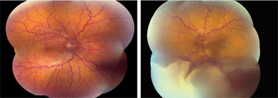 | |
| 1, 2. Color photo montage (O.D. left, O.S. right) shows obvious retinal changes in the left eye. Look carefully and you can see subtle changes in the right eye as well. |
Dilated fundus exam revealed small cups with good rim coloration and perfusion. Other changes can be seen in the fundus photos of both eyes (figures 1 and 2). A fluorescein angiogram is also provided (figures 3 and 4).
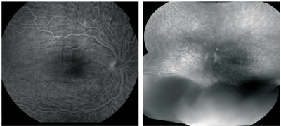 |
| 3, 4. Fluorescein angiography (O.D. left, O.S. right) shows changes in both eyes, but these were more obvious in the right eye. |
Take the Retina Quiz
1. What do the changes in the patients left eye likely represent?
a. Retinal detachment.
b. Choroidal detachment.
c. Combined choroidal and retinal detachments.
d. Choroidal melanoma.
2. What does the fluorescein angiogram of the patients right eye reveal?
a. Exudative retinal detachment.
b. Choroidal detachment.
c. Choroidal folds.
d. Optic nerve edema.
3. What is the correct diagnosis?
a. Metastatic carcinoma.
b. Uveal effusion syndrome.
c. Vogt-Koyanagi-Harada (VKH) syndrome.
d. Choroidal melanoma.
4. How would you characterize the overall condition of her eyes?
a. Microphthalmic.
b. Anophthalmic.
c. Nanophthalmic.
d. Buphthalmic.
5. What laser treatment did she likely have?
a. LASIK.
b. Peripheral laser photocoagulation.
c. Laser peripheral iridotomy.
d. Selective laser trabeculoplasty.
For answers, see below.
Discussion
The peripheral retinal changes in the left eye represent combined retinal and choroidal detachments. Although we can only see the choroidal detachments inferiorly, they extend circumferentially 360 degrees. We did not observe choroidal detachments in the right eye; however, there is serous fluid around the optic nerve that you can just make out on the fundus photo.
Also of note: Choroidal folds, seen on the fluorescein angiogram, were present in both eyes but were more prominent in the right eye. These show up on fluorescein angiography as alternating light and dark radial lines that extend above and below the macula.
So, what is going on with our patient? Besides the choroidal detachments, the high amount of hyperopia provides an important clue. She also has a small axial length of about 17mm in both eyes. The small axial length and high hyperopia makes this a nanophthalmic eye.
The headache may have been from her high IOP or from inflammation. The black spots and silver rings may be from the detachments and fluid under her retina, particularly in the macula.
All these findings indicate that our patient has uveal effusion syndrome. Uveal effusion syndrome represents a disease process in which patients develop serous detachments of the choroid, ciliary body and eventually the retina. It most often occurs in healthy middle-aged males, but it can occur with either sex. There is also a relationship between uveal effusion syndrome and nanophthalmic eyes.1
Patients who have uveal effusion syndrome most often present with loss of vision in one or both eyes due to serous retinal detachment involving the macula. They may also present with loss of peripheral vision secondary to choroidal and/or retinal detachments. The anterior segment exam of these patients is unremarkable, with no signs of inflammation, and the vitreous may show occasional cells.
These are important and noteworthy features, as Vogt-Koyanagi-Harada syndrome (VKH) can also present in a similar fashion. Patients who have VKH, however, exhibit inflammation in both the anterior segment and vitreous.
An important feature of uveal effusion syndrome is that there may be marked shifting of the subretinal fluid as the patient moves his or her eyes or lies in a supine position. This is due to a high protein content of the serous fluid, which is two-and-a-half to three times higher than normal plasma. The shifting of serous fluid is not seen in patients that have retinal detachment secondary to retinal tears or holes.
The pathogenesis of uveal effusion syndrome is poorly understood. It seems to occur in patients who have unusually thick scleras, as seen in nanophthalmic eyes. The abnormally thick sclera can compress the vortex veins and impede the venous drainage.2 Another theory is that the thick sclera may represent a barrier to the bulk fluid flow out of the eye.2 In these cases, surgical thinning of the sclera has been shown to resolve the detachment. Uveal effusion can also occur in eyes that are not nanophthalmic but have a thick sclera.
The treatment of uveal effusion syndrome can be complicated. As mentioned, surgical thinning of the sclera has been shown to be successful. Other approaches include segmental or partial-thickness sclerectomies and scleral decompression of the vortex veins.
Our patient underwent scleral decompression of all four vortex veins plus a pars plana vitrectomy with a gas fluid exchange and membrane peel. Despite the surgery, she continued to have persistent fluid that would not resolve. She underwent a second surgery but still had persistent retinal detachments. She continues to be followed closely.
Retina Quiz Answers: 1) c; 2) c; 3) b; 4) c; 5) c.
1. Gass JDM. Stereoscopic Atlas of Macular Disease. Idiopathic Uveal Effusion Syndrome. 4th ed. St. Louis: CV Mosby Co., 1997:200-5.
2. Bird AC. Pathogenesis of serous detachments of the retina and pigment epithelium. In: Ryan SJ. Schachat AP, Murphy RP, eds. Retina, Vol. IIMedical Retina. 4th ed. St. Louis: CV Mosby Co., 2005:974-5.
