 |
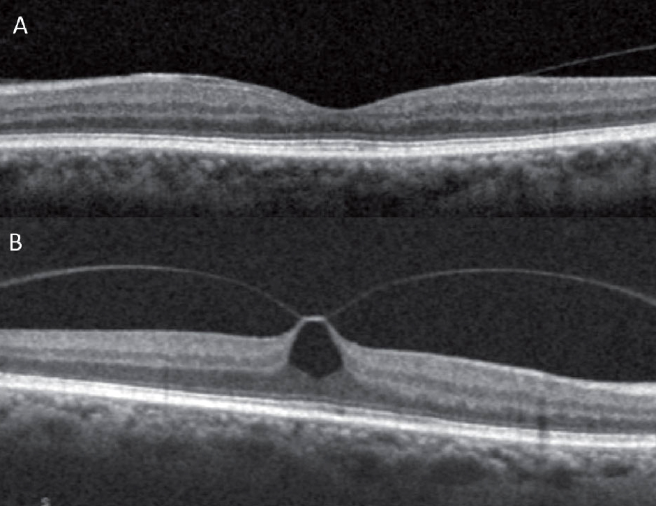
| The researchers suggested that future studies can be conducted prospectively to analyze the progression of VMA (fig. A) as a means of anticipating vitreomacular traction (fig. B) and subsequent macular hole development. Photo: Anna Bedwell, OD. Click image to enlarge. |
One of the more common sequelae of posterior vitreous detachment, vitreomacular adhesion (VMA) describes residual attachment between the vitreous and macula due to incomplete separation. Although it might not lead to any immediate retinal abnormality, VMA may exert traction on the underlying macula, distorting the retinal architecture and leading to vitreomacular traction. It may also contribute to the development of various retinal complications. Researchers in India decided to analyze the distribution of SD-OCT biomarkers in different types of VMA and the associated visual impairment in diabetic macular edema (DME). They found a similar prevalence of biomarkers in eyes with or without VMA, but the presence of these biomarkers was more likely to be associated with visual impairment in eyes with VMA. The team recommended that clinicians schedule regular follow-ups, especially monthly SD-OCT, if available, to specifically evaluate the vitreomacular interface in patients with DME.
A total of 317 eyes of 202 patients were enrolled. Cases were divided into two groups focal VMA (n=103) and broad VMA (n=104) and subjects with no VMA (n=110) were enrolled as controls.
The central retinal thickness and central subfield thickness values were statistically significant between the groups. Except for inner nuclear layer cysts, absence of bridging tissue that is composed of muller cell fibers and bipolar cells and hyper reflective dots in cyst, there were no significant differences in the distribution of OCT biomarkers among the three groups. Only disorganization of retinal inner layers showed significant association with vision impairment in all the three groups.
“The study results demonstrate that the distribution of OCT biomarkers is similar among all three groups. Still, the visual impairment was largely identified in subjects with a broad VMA group than the other groups,” the researchers wrote in their paper. “Vitreoretinal interface abnormalities play an important role in pathophysiology of macular disease.”
The team highlighted that SD-OCT “emerges as a clinically valuable and readily available method that can identify specific abnormalities at the vitreomacular interface that might not be detectable using ultrasonography or indirect ophthalmoscopy.”
To ensure early detection and effective management of subsequent visual complications in patients with DME, “it is advisable to closely monitor VMA in the long term,” they concluded.
Subramanian B, Devishamani C, Raman R, Ratra D. Association of OCT biomarkers and visual impairment in patients with diabetic macular edema with vitreomacular adhesion. PLoS One. 2023;18(7):e0288879. |
