Millions of Americans have had corneal refractive surgery. As these people age, they are now beginning to develop cataracts. These are patients who previously took steps to reduce the use of optical devices, so their postoperative cataract surgery expectations are typically much higher than those of the average patient.
Several factors make it much more difficult to achieve an accurate refractive outcome for postoperative corneal refractive patients. The main problem: Measuring the corneal power is difficult after refractive surgery, so calculating the power of the intraocular lens is much more complex in these cases.
Optometrists must understand what makes these post-refractive surgery calculations difficult so they can take steps to aid postoperative refractive patients in obtaining the information they will need when they develop cataracts. Having this information will make it easier for the patients cataract surgeon to achieve a more accurate calculation of the patients IOL power.
A Growing Problem
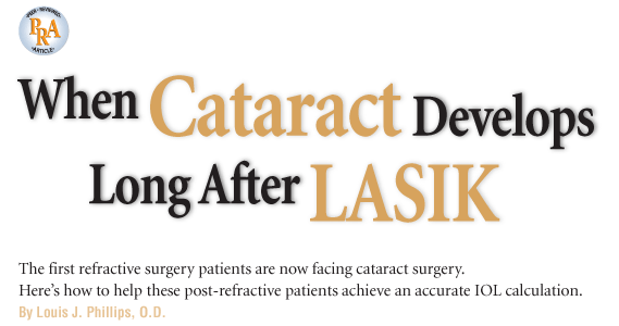
This map shows the patients preoperative topography with simulated K, which is very close to the manual K.
Since the Food and Drug Administration approved LASIK in late 1995, more than 7.1 million Americans have had the procedure, according to the research firm Market Scope LLC. The average age of the laser patient has consistently remained around 39 years old.1 This means that the average age of patients who had refractive surgery in 1996 is now 51, with many in their 60s.
In small but ever-growing numbers, these patients are arriving for cataract surgery. Obviously, larger numbers of these patients will need cataract surgery in the future, so we all will have more of these patients in our practices.
Calculating IOL Power
The calculation of the IOL power for cataract surgery requires the accurate measurement of the corneal power, axial length, anterior chamber depth and corneal diameter of the eye. These measurements are then plugged into formulae that calculate the power of the IOL needed to achieve the desired refractive outcome. These formulae have been developed over time and have reached such a level of sophistication that they are now even able to estimate the position of the implant in the eye to more accurately calculate the power of the IOL.2
In a postoperative refractive patient, the traditional measurements of the corneal power are no longer correct. This inaccuracy in corneal power measurement also throws off the estimation of the IOL position in the eye, which compounds the IOL
power error. If no adjustments are made for this corneal error, the calculation of the IOL power will be inaccurate by an amount directly proportional to the level of the refractive surgery correction.
Standard instruments that measure the corneal power actually measure the radius of curvature of the anterior surface, and then assume an index of refraction of the corneal tissue and a relationship of the anterior surface to the posterior surface. This is the information used to determine the refractive power of the cornea.
Laser refractive surgery does not change the index of refraction, but it does change the relationship of the anterior corneal surface to the posterior corneal surface. To achieve a diopter of myopia refractive correction with laser surgery, the anterior surface is flattened by approximately 1.4mm. As measured by keratometry, this would equal approximately 0.75D less power. Thus, for every diopter of correction achieved by laser surgery, the measured corneal power only changes by three quarters of a diopter. Therefore, corneal power measurements taken with standard instruments subsequent to myopic laser refractive surgery are measured stronger than the true corneal power. If this error in corneal power is inserted into the standard formulae, the IOL chosen will result in a hyperopic outcome.
For example, if a patient with a corneal power of 44.00D preoperatively has 4.00D of myopia treatment at the corneal plane; the true corneal power will then be 40.00D. The measured power, however, will be 41.00D. If the unadjusted measured power is used in calculation of the IOL power for a cataract patient, the outcome will be 1.00D of hyperopia. This occurs because the formula uses a measured corneal power that is 1.00D stronger than the actual power. Similarly, if a patient with the same corneal power has 8.00D of treatment at the corneal plane, then the true corneal power will be 36.00D but the measured power will be 38.00D, causing a 2.00D hyperopic error.
Correct K Readings
As the above discussion suggests, we must adjust the measured corneal power to arrive at a more accurate estimate of the true corneal power by using either the preoperative corneal power measurement plus the achieved refractive correction at the corneal plane or the postoperative corneal power measurement minus approximately 25% of the achieved refractive correction.
In the preoperative method, the achieved refractive correction is subtracted from the preoperative Ks. This requires accurate preoperative K readings and an accurate stable postoperative refraction.
Preoperative K method
Measured preoperative K: 44.00D
Achieved refractive correction at the corneal plane: -8.00D
True corneal refractive power = Preoperative K + achieved refractive correction
36.00D = 44.00D + (-8.00D)
In the postoperative method, one quarter of the achieved refractive correction is subtracted from the measured postoperative K reading.
Postoperative K method
Measured postoperative K: 38.00D
Achieved refractive correction at the corneal plane: -8.00D
True corneal refractive power = Postoperative K + (0.25 x -8.00D)
36.00D = 38.00D + (0.25 x -8.00D)
Both of these methods have inherent problems that can cause inaccuracies:
A careful K reading is often not taken preoperatively because it does not impact the refractive surgery correction.
An accurate postoperative refraction often is not done, especially if the patient has reasonably good uncorrected acuity and no vision complaints.
Accurate measurement of the postoperative corneal curvature with many of the instruments available is difficult. For that reason, we use several instruments including manual keratometer, topographer, Orbscan (Bausch & Lomb) and IOLMaster (Carl Zeiss Meditec) to measure the postoperative curvature of the cornea and evaluate all findings, usually giving more weight to the manual K reading.
Another confounding factor for both of these methods is that corneal curvature may change over time after refractive surgery. This is not a frequent problem, but minor ectasia following LASIK or epithelial hyperplasia following PRK will throw these calculations off.
Both methods require historical records. Yet it is often difficult, even today, to locate patient records. We have had doctors digging for records in their office basements and patients digging through their junk drawers to find old glasses or contact lenses for any piece of information that may help in the calculation.
Some years ago, we saw one patient who underwent LASIK in
In this case, it was fortunate that the patient could accurately assist in estimating his preoperative refractive error, but many patients cannot. This situation is going to get worse. Can you imagine trying to find a 20- or 25-year-old record, when locating even a 10-year-old record is causing us problems?
Establishing an accurate corneal power is the most important task in arriving at a good refractive outcome for these patients. However, another important step must be taken. Modern IOL calculation formulae predict the postoperative position of the IOL in the eye. This adjusts for the effect of vertex distance. In the postoperative refractive surgery eye, the corneal shape is changed but the internal structures of the eye are not. As a result, these formulae calculate an inaccurate lens position, which introduces another error factor.
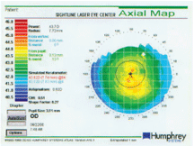
This map shows the patients postoperative topography simulated K which is different than the manual K (38.62 x 38.87=SE38.75).
Postoperative refractive surgery formulae have been developed to adjust for this error.3-6 These formulae are used to calculate IOL powers for these patients. The American Society of Cataract and Refractive Surgeons (ASCRS) has established a Web site calculator containing postoperative refractive surgery formulae. Doctors can input data for each formula to calculate the IOL powers (http://iol.ascrs.org). Accurate historical data are still needed.
You Can Make a Difference
Until instruments are developed that can accurately measure the postoperative anterior and posterior corneal curvature and calculate the true corneal power, we will need historical information. Instruments, such as the Pentacam (Oculus), have been developed that are getting better at measuring true corneal power in post-refractive surgery patients. Even when we reach this goal, we still need the achieved refractive correction to accurately estimate the proper IOL position in the eye.
It is important to inform any corneal refractive patient of the significance of this pre- and postoperative information. The second step is to make the information available to them.
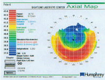
The numerical view of the same patient shows an average of the 0mm to 4mm ratings of 38.83, which is much closer to the manual K. This demonstrates the kind of problem encountered with various methods of measuring postoperative corneal curvature. Effective corneal power calculates to ~37.37 diopters for both the pre-K and post-K methods for this -6.37 refractive treatment.
To that end, a number of years ago in my private practice, we designed a card that contains the needed pre- and postoperative information. We give all of our corneal refractive patients a completed copy of this card at their three-month visit. This card is made of heavy paper stock, and we instruct patients to put it with their birth certificate and other important papers so it will be available if and when they need it. The card includes the patients preoperative and postoperative refractions, keratometry readings and IOP.
A preoperative K reading needs to be accurate and, ideally, should be a manual K reading. This K reading should be taken after corneal stabilization if the patient wears contact lenses. An accurate preoperative refraction is found in most records because it is important for an accurate outcome for the refractive surgery. The postoperative K reading and refraction are measured and recorded as accurately as possible at the three-month visit.
Pre- and postoperative findings are easy to collect if the patient starts with you. It is more difficult to help a patient who comes to you after refractive surgery has been done. It is more likely that the patient can find the preoperative information now than in the future. Give the patient the Refractive Surgery Card or a prescription pad listing the needed information and instruct them to contact their previous practitioner immediately to acquire the information. Or, you can ask the patient to sign a records release so you can obtain the information for them.
The other information that is valuable for future practitioners is the pre- and postoperative intraocular pressure. It is not needed for lens calculation, but when the cornea is thinned by refractive surgery, the measured IOP changes. Depending on how much the cornea is thinned, the postoperative measured intraocular pressure can be lowered by several millimeters of mercury (mm Hg).
The use of this card demonstrates to your patients that you care about their future vision needs. This is especially true if you seek them out to give them this information. Many postoperative refractive patients do not return for routine care. If you can identify past refractive surgery patients in your practice through your computer system, you can send them a completed Refractive Surgery Card along with a reminder of their need for ongoing care. This goodwill gesture can help retain patients and spur referrals.
Most importantly, though, making this information available will help your patients in the future.
When the History is History
A number of regression formulae have been developed to calculate the IOL power for patients when no historical information is available.7 These formulae use the postoperative K measurement and an adjustment based on collected data from refractive surgery patients. These formulae are still a less accurate way of calculating the post refractive surgery IOL power than the formulae that use the historical data.
Supplying this information to patients is not an easy task, and in some cases, it will be impossible. It is important enough, however, to justify the significant effort it will take. If you start to make this information available to your old and new laser refractive patients now, youll be delivering a service that will be valuable to them in years to come.
Dr. Phillips is president of the
1. Personal correspondence with Dave Harmon, Market Scope, LLC.
2. Narvaez J, Zimmerman G, Stulting RD, Chang DH. Accuracy of intraocular lens power prediction using the Hoffer Q, Holladay 1,
3. Walter KA, Gagnon MR, Hoopes PC Jr, Dickinson PJ. Accurate intraocular lens power calculation after myopic laser in situ keratomileusis, bypassing corneal power. J Cataract Refract Surg 2006 Mar;32(3):425-9.
4. Aramberri J. Intraocular lens power calculation after corneal refractive surgery: double-K method. J Cataract Refract Surg 2003 Nov;29(11):2063-8.
5. Latkany RA,
6. Fam HB, Lim KL. A comparative analysis of intraocular lens power calculation methods after myopic excimer laser surgery. J Refract Surg 2008 Apr;24(4):355-60.
7. Shammas HJ, Shammas MC. No-history method of intraocular lens power calculation for cataract surgery after myopic laser in situ keratomileusis. J Cataract Refract Surg 2007 Jan;33(1):31-6.
