 An 11-year-old white female presented with reduced acuity in her left eye that had persisted for six weeks. She denied any ocular trauma or other insult that would have contributed to her blurred vision.
She had never worn glasses. In fact, the patient never had an eye examination in her life. Her medical history was unremarkable. She did not take any medications and had no known allergies.
On examination, her best-corrected visual acuity measured 20/20 O.D. and 20/200 O.S. Confrontation visual fields were full to careful finger counting O.U., and her extraocular motility testing was normal. Her pupils were equally round and reactive, with no afferent defect. Her anterior segment examination was unremarkable. Her intraocular pressure measured 15mm Hg O.U.
The dilated fundus exam of her right eye was completely normal. However, the fundus exam of her left eye revealed an obvious yellow-white lesion adjacent to the optic nerve that extended superiorly (figure 1). On optical coherence tomography, we also noted some changes in her left macula (figure 2).
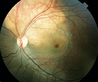
1. A fundus photograph of our patient’s left eye at
her initial office visit. Note the yellow-white change located adjacent
to her optic nerve superiorly. What does this change represent?
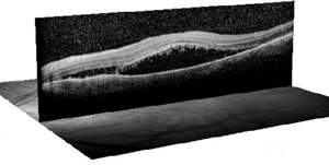
2. An OCT image through her left macula. What does this scan reveal?
Take the Retina Quiz
1. What does the yellow-white lesion represent?
a. Retinoblastoma.
b. Amelanotic melanoma.
c. Choroidal osteoma.
d. Choroidal hemangioma.
2. What additional testing would be most useful?
a. Standardized ultrasound.
b. Fluorescein angiogram (FA).
c. Magnetic resonance imaging (MRI).
d. Fine needle biopsy.
3. What do the changes in the macula represent?
a. Serous detachment.
b. Retinal pigment epithelium (RPE) detachment.
c. Cystoid macular edema (CME).
d. Choroidal and RPE detachment.
4. What does the OCT of the macula show?
a. Serous detachment.
b. RPE detachment.
c. CME.
d. Choroidal and RPE detachment.
5. How should this patient be managed?
a. Close observation.
b. Laser treatment.
c. Anti-VEGF therapy.
d. Enucleation.
For answers, see below.
Discussion
Our patient has a choroidal osteoma with a secondary serous retinal detachment involving her macula. A choroidal osteoma is a benign tumor of the choroid that is comprised of mature bone. Most commonly, it occurs in healthy, young females aged 10 to 20 years. Most chorodial osteomas have been reported in whites; however, a few cases have been observed in patients of other races.1
A choroidal osteoma classically presents as a yellow-white to orange-red lesion that is located in the juxtapapillary or peripapillary region. The exact coloration and appearance of a choroidal osteoma can vary depending on the extent of RPE thinning or depigmentation. These lesions can range from 0.5mm to 2.5mm in elevation, with an irregular surface due to natural variations in the osseous tumors. These tumors have a vascular system, and their blood vessels may be especially visible in areas where the overlying RPE is thin or depigmented. And, because the vessels can be large, they are often mistaken for CNV.1
Approximately 75% of choroidal osteoma cases present unilaterally and asymptomatically. The majority of tumors are often discovered as an incidental finding during a routine eye examination. Most osteomas grow slowly over a period of months to years. However, there have been reports of tumors doubling in size as well as new tumor growth in healthy, previously examined eyes.1
One of the most common complications of choroidal osteomas is the development of subretinal serous fluid that overlies the lesion. Also, choroidal neovascularization has been shown to occur in up to 47% of eyes over a 10-year period, and up to 57% after 20 years.1,2
Indeed, our patient had a serous detachment overlying the tumor that was visible on OCT. Interestingly, there was also a schisis cavity present between the osteoma and the serous detachment that involved her macula. This schisis may explain why the serous fluid overlying the tumor extended into her macular region. As the fluid increased, it extended into her macula, which resulted in symptomatic vision loss.
So, how is it possible for a bony tumor to present in the eye? The exact pathogenesis is unknown, but hormonal influences may play a role, as evidenced by the condition’s predilection for young females.1 Nonetheless, it is believed that ossification occurs wherever there is an abundant blood supply that can support the architecture of the Haversian system, a series of canals or tubes that surround the narrow channels formed by lamellae. These lamellae surround blood vessels and nerve cells throughout the bone. The osteoma is composed of dense bony trabeculae with large, endothelial-lined cavernous spaces and small capillary blood vessels. Osteoblasts, osteocytes and osteoclasts may also be present.1
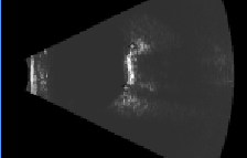
3. Our patient’s B-scan showed a slightly elevated, highly reflective choroidal mass.
Ultrasound is likely the best diagnostic test to differentiate an osteoma from a similar lesion. The B-scan will show a highly reflective mass that persists at lower sensitivities with acoustic shadowing of the orbit posterior to the mass. These features were present in our patient (figure 3).
Our patient was completely asymptomatic until she developed the serous detachment involving her left macula. We performed an FA, which showed diffuse leakage but no CNV. Initially, we elected to closely observe the patient to see if the serous detachment would self-resolve. Unfortunately, the detachment enlarged significantly within two weeks of her initial office visit. So, we administered an intravitreal injection of Avastin (bevacizumab, Genentech) while she was under anesthesia. We will continue to follow her progress during the next several weeks.
Answers to the Retina Quiz: 1) c; 2) a; 3) a; 4) a; 5) a.
1. Shields, CL, Brown GC, Sharma S, et al. Choroidal Osteoma. In: Ryan SJ, Schachat AP, Murphy RP (eds). Retina, Vol. III: Chapter 46, Tumors of the Retina, Vitreous and Choroid, 4th ed. St. Louis: Mosby, 2006:819-28.
2. Aylward GW, Chang TS, Pautler SE, et al. A long-term follow-up of choroidal osteoma. Arch Ophthalmol. 1998 Oct;116(10):1337-41.
