 |
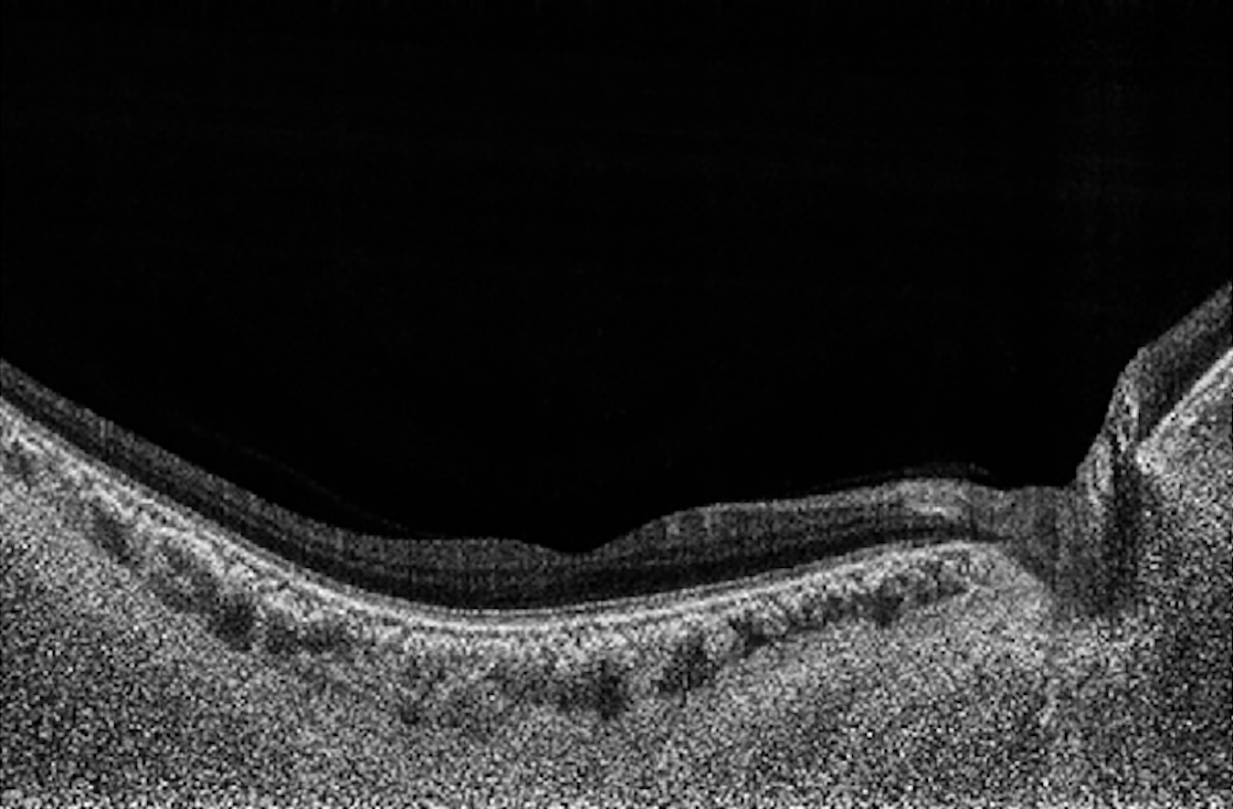
3D widefield OCT scans appear to detect structural glaucomatous damage better than traditional scans of the optic nerve head or macula, study finds. Photo: Chiang CYN, et al. Transl Vis Sci Technol. 2024;13(1):5. Click image to enlarge. |
When managing patients with primary open-angle glaucoma, optometrists must be vigilant in monitoring and identifying structural changes in the macula and optic nerve head (ONH), as early detection of damage allows for sooner intervention to stop disease progression and preserve vision. The recent innovation of three-dimensional widefield OCT means that doctors can now monitor both the macula and ONH at once, but does this method improve POAG diagnosis? A recent study says yes; it concluded that examining both structures in tandem via 3D widefield imaging had greater diagnostic power than traditional ONH or macula OCT scans.
Researchers started by developing a deep-learning algorithm to automatically segment the ONH and macula structures in 3D-OCT scans. The algorithm was able to automatically label major tissue structures, trained with 270 manually annotated B-scans. Performance was assessed using the Dice coefficient. Next, the team designed a glaucoma classification algorithm using 500 OCT volumes and corresponding automatically segmented labels. This algorithm was then trained and tested on three datasets: cropped scans of macular tissues, those of ONH tissues and widefield scans.
After comparing 319 OCT scans of glaucoma eyes and 298 of non-glaucoma eyes, the researchers reported that the ONH and macula structures were able to be accurately segmented using the deep-learning algorithm, which achieved a Dice coefficient of 0.94. They wrote in their paper on the study findings, published in Translational Vision Science & Technology, “The classification algorithm was best able to diagnose glaucoma using widefield scans, followed by ONH scans, and finally macula scans, with AUCs of 0.99, 0.93 and 0.91, respectively.”
Widefield imaging may be more accurate for glaucoma diagnosis than ONH or macula scans alone due to the additional information it provides that adds value to the POAG classification process, the researchers explained. Similarly, the superior performance of ONH scans over macula scans suggests that the ONH region on widefield OCT may provide more data than the macula region for classifying POAG vs. non-POAG, they added. Because of this, the authors noted in their paper that “ONH OCT scans, instead of macula OCT scans, should be preferred for glaucoma studies using AI or deep learning where widefield scans are not available.”
Given this new evidence that widefield imaging can improve POAG diagnosis over traditional OCT scans, this may encourage more ODs to incorporate the technology into their practices, as most OCT devices now offer this modality, the researchers concluded.
Chiang CYN, Braeu FA, Chuangsuwanich T, et al. Are macula or optic nerve head structures better at diagnosing glaucoma? An answer using artificial intelligence and wide-field optical coherence tomography. Transl Vis Sci Technol. 2024;13(1):5. |
