The precision of OCT imaging is easily undermined by various factors that can lead to data artifacts, such as media opacity, misalignment, motion blur and several others. Refractive error, and astigmatism in particular, has been less well studied as a source of erroneous findings in OCT angiography (OCT-A), so a team of Spanish researchers investigated its influence. They found that astigmatism of -2D affects quantification of vessel density, mainly in the superficial vascular complex.
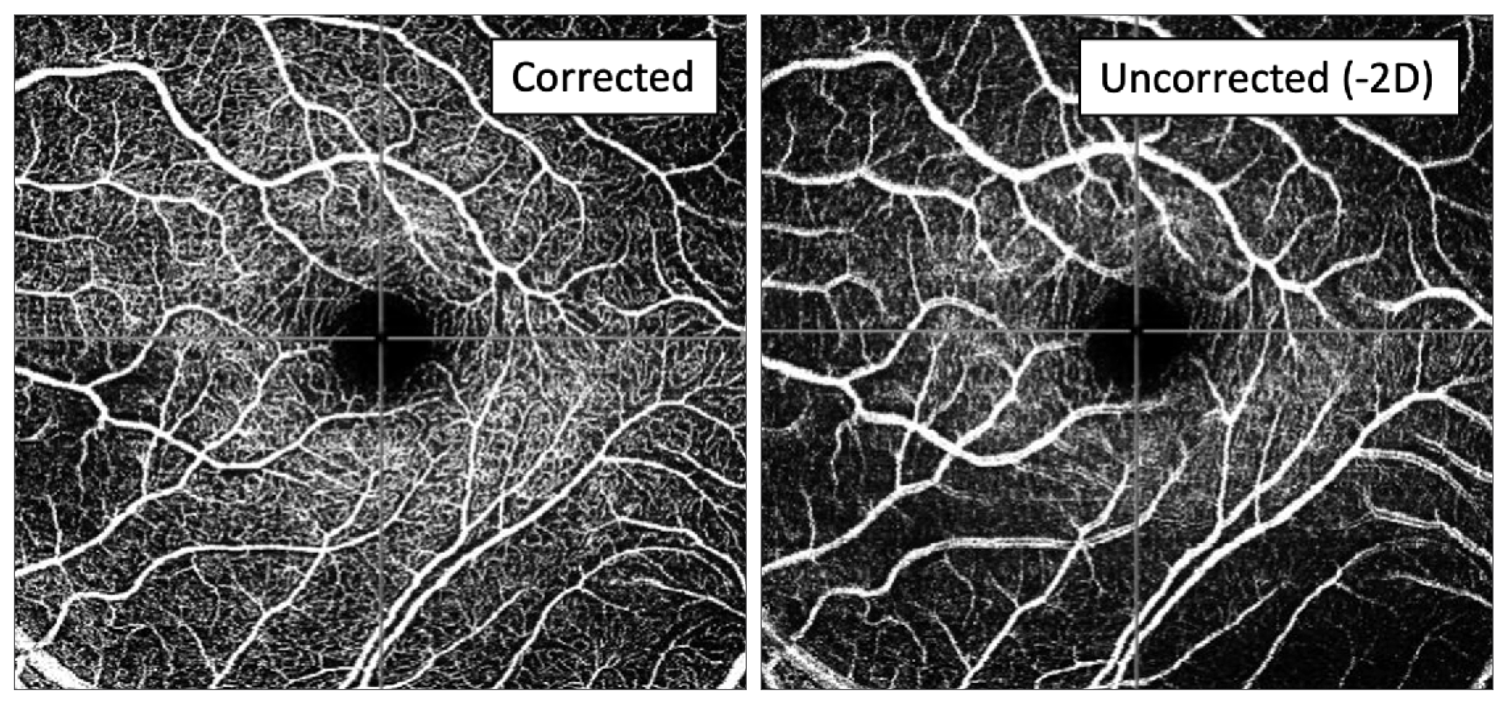 |
| OCT-A of the superficial vascular plexus with and without correction of -2D cyl shows less definition of capillaries and increased diameter of large retinal vessels induced by astigmatism. Photo: Vidal-Oliver L, et al. Transl Vis Sci Technol. Jan 15, 2024. Click image to enlarge. |
A total of 110 eyes were included: 20 without astigmatism and 90 with a half-diopter or more of astigmatism. In patients without astigmatism, follow-up scans were performed after induction of -1D and -2D cyl. In those with astigmatism, the follow-up scan was performed after astigmatism correction. A set of cylindrical lenses was attached to the camera head of the Spectralis. A quantitative and qualitative analysis of the superficial vascular complex and deep vascular complex was performed. The main outcome measures were vessel density, image quality and the presence of artifacts.
|
To learn more about OCT technology and download a handout that describes imaging artifacts, see this feature. |
Two diopters of astigmatism significantly affected vessel density measurements, as well as caused qualitative changes in OCT-A en face images, mostly in the superficial vascular complex. The degree of astigmatism might increase the vessel density dropout, the authors suggested.
“These changes might not be clinically meaningful, as they change in order of 0.012 to 0.02 in a per-diopter basis,” the authors noted in their paper for Translational Vision Science & Technology. “However, it could be relevant in studies addressing quantification changes, and should be considered as a limiting factor.”
They also found similar effects in the quantitative metrics after inducing astigmatism. The influence of astigmatism was weaker in the deep vascular complex but showed similar trends compared to the effect on the superficial vascular complex.
“We hypothesized that other artifacts may play a role in this deep slab, and that differences in vessel density calculations on capillary vessels might be more difficult to assess,” the authors explained. “In addition, we found that the differences found in the superficial vascular complex were significant in more areas compared with the deep vascular complex. The superficial plexus is the least affected by projection artifacts, which are usually present in deeper layers, such as the intermediate and deep plexus.”
The qualitative assessment showed worse image quality in the images acquired in the presence of astigmatism (astigmatic induction in patients without astigmatism, mostly of -2D, or corrected images in those whose astigmatic error was higher or equal to -0.50D), both in the superficial vascular complex and the deep vascular complex.
“In addition, the percentages of images showing attenuation and defocus artifacts were higher in those images acquired with astigmatism, which can also explain the vascular dropout demonstrated in the quantitative analysis,” the authors pointed out.
“In summary, we showed that the presence of -2D of astigmatism resulted in significantly reduced VD measurements, as well as diminished image quality including large vessel and capillary blurring. This refractive error must be considered in studies addressing quantitative OCTA parameters. Further studies with a higher number of patients and a wider range of astigmatic defects are needed to create a more accurate model to estimate VD changes in relation with astigmatism diopters.”
Vidal-Oliver L, Pinazo-Gallego R, Dolz-Marco R. Astigmatism influences quantitative and qualitative analysis in OCT angiography imaging. Transl Vis Sci Technol. Jan 15, 2024. [Epub ahead of print.] |
