A recent study provided evidence of variance between geographic atrophy (GA) characteristics in Asian vs. Caucasian patients, which could potentially have implications when determining the most effective course of treatment. After conducting a retrospective multicenter observational study on a Japanese cohort with GA associated with age-related macular degeneration (AMD), researchers identified several clinical differences, such as male dominance and a relatively thicker choroid in Asian vs. Caucasian patients with GA. The Asian population also had small lesions and a lower disease progression rate.
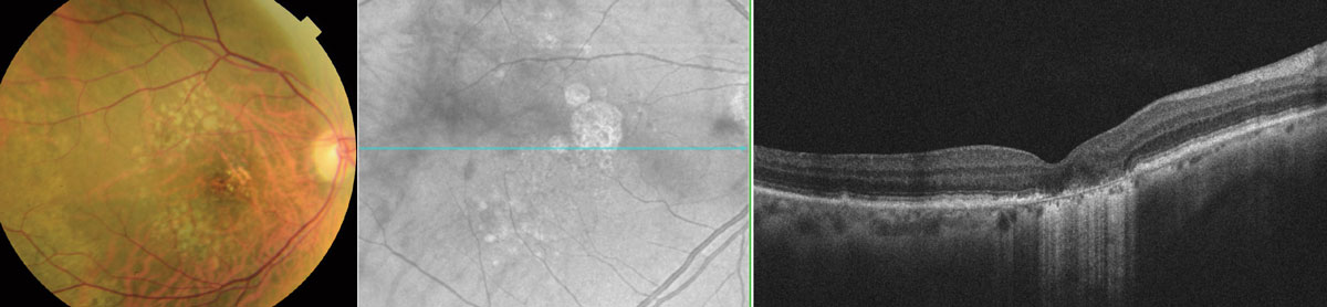 |
| Japanese patients in this study had a smaller GA lesion size than previously reported in Caucasians (306mm2 vs. 4.62mm2), as well as slower GA progression (1.01mm2 vs. 1.27mm2). Photo: Wendy Harrison, OD, PhD. Click image to enlarge. |
A total of 173 eyes of 173 patients from six university hospitals in Japan were included in the study. Of those, 101 eyes of 101 patients were included in the follow-up group. All patients were at least 50 years old and had definite GA associated with AMD in at least one eye. The mean age was 76.8, and 63% of the cohort was male. To measure the GA area, researchers used fundus autofluorescence.
The mean GA area was 3.06mm2, and more than a third of patients (35.8%) had bilateral disease. The researchers reported the following additional clinical characteristics of the Asian cohort:
- Twenty-two percent of eyes were classified as having pachychoroid GA.
- Drusen and reticular pseudodrusen were detected in 66.5% and 42.2% of eyes, respectively.
- The mean subfoveal choroidal thickness was 194.7µm.
All patients in the follow-up group showed progression of GA over time (average follow-up: 46.2 months). The mean progression rate was 1.01mm2/year, which is less than what has been reported in Caucasian populations (1.27mm2/year). A multivariable analysis revealed that a greater GA progression rate was significantly associated with a larger baseline GA area and the presence of reticular pseudodrusen.
The researchers summarized the findings in their paper for Ophthalmology Retina, noting that “GA in this Asian population was male-dominant, had small lesions, a relatively thicker choroid and a low GA progression rate. There was a group with GA without drusen but with features of pachychoroid. In addition, large baseline GA area and reticular pseudodrusen were associated with faster GA progression.”
Prior research has observed some differences in GA characteristics of Asian vs. Caucasian populations, the researchers pointed out. “Previous studies on Caucasian patients showed no sex differences in the prevalence of GA; however, in our study, patients with GA were predominantly males (63%),” they wrote in their paper. “This tendency is consistent with a meta-analysis on GA in Asian populations, which showed a prevalence of 1.62/1,000 in males and 0.87/1,000 in females.”
The GA size observed in Japanese patients in this study was also smaller than that reported in previous similarly designed studies on Caucasians (306mm2 vs. 4.62mm2).
The researchers concluded that their findings “indicate that some characteristics of Asian patients with GA differ from those of Caucasians. Researchers need to consider the differences in the phenotype of GA in different ethnicities as these differences may have implications when researching GA and considering interventions to slow the progression of GA.”
Sato Y, Ueda-Arakawa N, Takahashi A, et al. Clinical characteristics and progression of geographic atrophy in a Japanese population. Ophthalmol Retina. June 5, 2023. [Epub ahead of print]. |
