 History
A 33-year-old black male presented for an annual eye examination. He had no complaints, and his ocular and systemic histories were unremarkable. He denied using any medications, and reported no known allergies.
Diagnostic Data
His best-uncorrected visual acuity measured 20/20 O.U. at distance and near. Refraction uncovered clinical emmetropia. His pupils were equally round, with no evidence of afferent defect.
Extraocular muscle movements were full and unrestricted in all positions of gaze. Confrontational fields were full O.U.
Slit lamp examination revealed normal and healthy anterior segment structures. His intraocular pressure measured 16mm Hg O.D. and 15mm Hg O.S. Dilated fundus examination uncovered normal posterior poles, with healthy and intact peripheries.
However, during gross external inspection, we observed the presence of a significant lesion (figure 1).
Your Diagnosis
How would you approach this case? Does this patient require any additional tests? What is your diagnosis? How would you manage this patient? What’s the likely prognosis?
Discussion
Additional testing should include a thorough history of the lesion. Be certain to ask the patient:
- How long has the lesion been present?
- How long has it appeared open and/or unhealed?
- Has another health care provider seen the lesion? If so, did he or she offer you some advice on how to proceed?
- Has the lesion changed size recently?
- Has the lesion changed color or elevation?
- Have you or any other family members previously had comparable lesions removed?
You should then photodocument the lesion. If a camera is not available, a hand-drawn illustration with physical measurements will suffice.
The patient in this case has basal cell carcinoma. BCC accounts for 75% of all skin cancers, with an incidence that continues to rise by 3% to 8% each year.1-3 Not only is BCC the most common form of skin cancer in humans, it also has the highest overall probability for malignancy.2,3
Typically, BBCs present as slow-growing, locally invasive epidermal skin lesions in older, light-skinned individuals.2,3
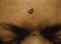
External examination of our patient revealed the presence of a significant lesion.
In most cases, the lesions develop secondary to chronic sun exposure. The most common locations for lesion development are the head and neck; lesions on the trunk and back are common as well. In addition to chronic ultraviolet radiation, other risk factors for the development of BCC include tanning bed use, a family history of skin cancer, lighter skin tone, a tendency to freckle in childhood, undergoing immunosuppression therapy, a history of previous radiotherapy and chronic exposure to certain toxic substances (e.g., inorganic arsenic).3
There are numerous variations in the clinical presentation of BCC: nodular, ulcerated (as is the case in our patient), pigmented, sclerosing, superficial and fibroepithelioma of Pinkus.3 Each form exhibits specific clinical features, a varying histopathology and characteristic behavior patterns (e.g., aggressive vs. less aggressive).
If left untreated or inadequately treated, BCC may become invasive and locally destructive.3 Fortunately, however, BCC rarely metastasizes.1-3
A little more than a decade ago, BCCs were widely regarded as a molecular black box.4 The discovery of “aberrant” hedgehog signaling in patients with a genetic predisposition to develop BCC has resulted in a better understanding of the abnormalities that could precipitate the condition.4-6 The first hedgehog gene was discovered in Drosophila (the common fruit fly), and consequently was named as such because it resulted in the proliferation of embryo phenotypes that were covered with pointy denticles that resembled a hedgehog.
The hedgehog signaling pathway (a normal process) plays a key role in directing growth and patterning during embryonic development in vertebrates.5 It is required for the normal development of many structures, including the neural tube, axial skeleton, skin and hair.5 Its activity is reduced or absent in adult organisms.6
Erroneous activation of the pathway (often referred to in the literature as “aberrant signaling”) has been shown to be a factor in the formation of several human malignancies.6 More specifically, aberrant activation of the hedgehog pathway in adult tissue has been associated with the development of BCC, medulloblastoma, and a subset of pancreatic, gastrointestinal and other cancers.5 This discovery better illustrated how the erroneous activation of this signaling pathway in adulthood might incite the growth of malignant lesions.4-6 Most importantly, a Phase I human trial of a hedgehog inhibitor has shown significant progress in halting––and even reversing––the growth of these tumors.7
In addition to the use of radiation therapy, topical cytostatics and immunomodulators, several surgical techniques––including Mohs Micrographic Surgery, cryotherapy/cryosurgery, curettage and carbon dioxide laser therapy, electrodessication and photodynamic therapy––could be employed to remove the lesion following detection.1
We referred our patient to a plastic surgeon for standard excisional biopsy. A pathologist diagnosed the lesion as BCC. Fortunately, the excision was sufficient and no additional radiation or pharmaceutical treatment was required.
1. Skelton LA. The effective treatment of basal cell carcinoma. Br J Nurs. 2009 Mar 26-Apr 8;18(6):346, 348-50.
2. Bath-Hextall FJ, Perkins W, Bong J, et al. Interventions for basal cell carcinoma of the skin. Cochrane Database Syst Rev. 2007 Jan 24;(1):CD003412.
3. Buljan M, Bulat V, Situm M, et al. Variations in clinical presentation of basal cell carcinoma. Acta Clin Croat. 2008 Mar;47(1):25-30.
4. Kudchadkar R, Lewis K, Gonzalez R. Advances in the treatment of Basal cell carcinoma: Hedgehog inhibitors. Semin Oncol. 2012;39(2):139-44.
5. Tang JY, So PL, Epstein EH Jr. Novel Hedgehog pathway targets against basal cell carcinoma. Toxicol Appl Pharmacol. 2007;224(3):257-64.
6. Caro I, Low JA. The role of the hedgehog signaling pathway in the development of basal cell carcinoma and opportunities for treatment. Clin Cancer Res. 2010;16(13):3335-9.
7. Epstein EH. Basal cell carcinomas: attack of the hedgehog. Nat Rev Cancer. 2008;8(10):743-54.
