 In last month’s “
Therapeutic Review,” my friend and colleague Dr. Joe Sowka recounted his story of developing an unusual and visually significant cataract at the ripe old age of 47. His tale and overview of “milky” nuclear sclerosis was insightful and reminds us all of an important lesson: namely, that while cataracts are usually easy to recognize and diagnose, they sometimes can be associated with strange circumstances.
In last month’s “
Therapeutic Review,” my friend and colleague Dr. Joe Sowka recounted his story of developing an unusual and visually significant cataract at the ripe old age of 47. His tale and overview of “milky” nuclear sclerosis was insightful and reminds us all of an important lesson: namely, that while cataracts are usually easy to recognize and diagnose, they sometimes can be associated with strange circumstances.
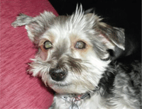
While atypical, Joe’s cataract certainly was not the most unusual case I have managed. In fact, I’ve had to go through bilateral cataract surgery with a member of my own household. No, it wasn’t one of my parents, my wife or even one of my children—it was my dog, Buddy.
Buddy is a Maltese-Yorkie mix (a “morkie”) that we found as a stray and adopted nearly two years ago. At the time, he was very healthy and playful, but after about a year, he became increasingly cautious, more fearful and less active. Then, he began bumping into doorjambs and missing steps. It was when he blindly walked into the pool, however, that we realized he had a significant vision problem that required examination.
Buddy’s Exam
Our veterinarian examined Buddy and confirmed what I already knew––he had dense bilateral cataracts. And although we have a pet insurance policy, cataracts are considered “elective surgery” and are not covered, even though hypermature canine cataracts often lead to lens-induced uveitis, secondary glaucoma and retinal detachment.1 So, Buddy’s doctor referred us to a veterinary ophthalmologist for consultation. At the specialist’s office, I watched in amazement as a technician performed a variety of very familiar tests on my dog. She visually inspected Buddy’s eyes, then instilled proparacaine and measured his intraocular pressure with a Tono-pen (Reichert). She stained his eyes with fluorescein and used a cobalt filter to check for corneal defects. She even performed a Schirmer test to measure his tear production. Then she instilled tropicamide, and we waited for dilation.
The surgeon performed biomicroscopy on Buddy using a portable, handheld slit lamp. She attempted binocular indirect ophthalmoscopy, but as we both soon realized, Buddy’s cataracts were too dense to permit a view of the fundus. We discussed the options, and I learned that cataract surgery for dogs is relatively common, although it is understandably more involved than in humans. Because animals must remain still and calm, general anesthesia is customary when performing ocular surgery. The lens is larger in dogs, and the lens capsule and cortex are thicker, making canine cataract surgery more challenging and protracted.2 Also, many people believe that intraocular lens implants are not required in animals because of their diminished visual demand, yet that is not always the case. In small dogs like Buddy (he weighs a mere 14 pounds), the lens accounts for 40.00D to 43.00D of the eye’s total power––in stark contrast to humans, where the lens power is about 16.00D to 18.00D.3 Without implants, surgery would have left my dog extremely hyperopic and vulnerable to injury or attack by larger animals.4
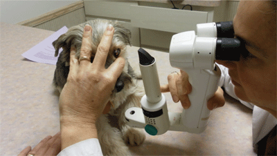
Christina Pellicane, D.V.M., performs biomicroscopy on Buddy prior to surgery.
Buddy’s Surgery
Agreeing that surgery was the right option, the veterinarian scheduled Buddy for additional testing that would include B-scan ultrasonography and a quantitative photopic and scotopic electroretinogram (ERG) to determine retinal function and evaluate for the possibility of progressive retinal atrophy.1 We also discussed whether to do surgery on one or both eyes.
While unilateral surgery is a less expensive option for restoring functional vision, there remains a strong risk of chronic, lens-induced uveitis and other complications in the unoperated eye.5 Conversely, bilateral surgery has the advantages of restoring sight with a single anesthetic episode and convalescent period, lower overall costs when compared with two unilateral surgeries, and a higher percentage of significant postoperative visual gain.1,6 We decided to pursue bilateral surgery.
The preoperative regimen was very similar to what we might perform in human patients, with one notable exception: Many of the medications used in veterinary medicine are what we would consider “older” drugs. For example, the antibiotic prescribed was not a fluoroquinolone, but rather a combination of neomycin, bacitracin and polymyxin (e.g., Neosporin ointment [Johnson & Johnson]). His NSAID was not nepafenac or bromfenac, but flurbiprofen. Additionally, Buddy was given oral amoxicillin and prednisone prior to surgery, which again, is not typical in human cataract surgery.
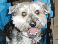
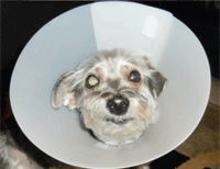
Buddy before surgery (top), at one-day post-op (middle) and six weeks post-op.
On the day of surgery, we dropped Buddy off in the early morning and he remained at the animal hospital until late that evening. This was necessary to ensure that he recovered adequately from the anesthesia and had no significant ocular or systemic complications. He was discharged with instructions to continue using all of the medications––including prednisolone acetate, tropicamide and multiple lubricants––and was given butorphanol tartrate to help sedate him. He also was fitted with an Elizabethan collar or “cone” to prevent him from instinctively rubbing at the sutures.
Buddy was doing well at his one-day post-op visit, save that his IOP was elevated. The veterinarian added two glaucoma medications—timolol and dorzolamide—to help address this issue. In all, Buddy remained on drops and in his cone for six weeks, until the incision line had vascularized and scarred over and his IOP normalized. Only one week after surgery, Buddy’s demeanor changed and he regained his confidence. Once again, he was jumping up on my furniture and running down the driveway to greet me when I came home from work.
While O.D.s rarely provide care for patients of the non-human variety, we are entrusted with public welfare and are viewed as a source of information on various eye care matters. For many patients, pets are part of the family. Their health is a primary concern, and owners will often seek the advice of their personal physicians in determining how to proceed in veterinary matters. So, a basic understanding of veterinary ophthalmology is a benefit for clinicians who engage in family practice optometry. Moreover, establishing a rapport with a local veterinary ophthalmologist, or even having their business cards to distribute when necessary, can help facilitate the care of these special four-legged patients.
Thanks to Christina Pellicane, D.V.M., and the Camelot Animal Hospital in Davie, Fla., for taking such great care of my little Buddy.
1. Wilkie DA, Colitz CM. Update on veterinary cataract surgery. Curr Opin Ophthalmol. 2009 Jan;20(1):61-8.
2. Warren C. Can your patient find the food bowl? Rev Ophthalmol. 2004 May;11(5):75-6.
3. Cataract surgery. Veterinarian Vision, Inc. Available at:
www.veterinaryvision.com/dvm_forum/dvm-cataracts.htm (accessed July 16, 2011).
4. Davidson MG. Refractive state of aphakic and pseudophakic eyes of dogs. Am J Vet Res. 1993 Jan;54(1):174-7.
5. Van der Woerdt A, Nasisse MP, Davidson MG. Lens-induced uveitis in dogs: 151 cases (1985–1990). J Am Vet Med Assoc. 1992 Sep 15;201(6):921-6.
6. Davidson MG, Nasisse MP, Rusnak IM, et al. Success rates of unilateral vs. bilateral cataract extraction in dogs. Vet Surg. 1990 May-Jun;19(3):232-6.
