 It was a strange scenario. A 32-year-old woman believed that her vision was getting blurry the evening before, but ignored it and went to bed. When she woke up the next day, her vision was “terrible.” After it failed to improve over several hours, she saw her internist emergently. He immediately referred her for a consultation.
Systemically, the patient was healthy and had no significant medical history. She used no current medications and denied any drug allergies. She had never worn glasses and rarely had her eyes examined, because her vision always had been “excellent.”
She was a healthy, well-nourished woman in no apparent physical distress. Her uncorrected visual acuity was 20/200 in each eye. Extraocular muscle testing showed no restrictions, and confrontation visual fields were normal in both eyes. Her eyelids were symmetrical, with no evidence of ptosis or eyelid retraction. However, of particular interest, we noted that she had bilaterally round, fixed and dilated pupils.
Neither pupil reacted to light or near accommodative stimuli. Her pupils measured 8.5mm OD and 9.0mm OS. Testing of the relative afferent visual system was impossible because of impaired pupillary reactivity.
Her fundus evaluation revealed normal optic discs and retinae OU. Interestingly (and tellingly), binocular indirect ophthalmoscopy was performed without the use of any mydriatic agents, because of the size of her dilated and unreactive pupils. Upon questioning, the patient denied trauma as well as use of any topical or systemic medications.
This presented quite the clinical conundrum. In this month’s column, we will describe how to evaluate patients who present with one or both pupils dilated.
Pupillary Evaluation
Evaluating pupils in both bright and dim light is vitally important. Further, pupils must be tested for response to both light stimulus (direct and consensual responses) and a near target (near synkinetic response). A history of pharmacologic use must be ascertained. A mydriatic agent will cause pupils to be round and unreactive to both light and near testing. A sympathetic agent will yield round, dilated pupils that likely will react––albeit sluggishly––to light and near stimulation.
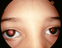
A dilated tonic pupil OD.
A history of trauma should be sought as well. Should the patient have experienced ocular trauma, carefully examine the iris via biomicroscopy for contributory signs, such as iridodialysis or pupil sector paralysis, because trauma can result in a fixed and dilated pupil. A famous example is singer David Bowie, who received a permanently fixed, dilated pupil at age 13 during a fight over a girl.
Typically, when encountering a dilated pupil, there will be some associated degree of anisocoria. When encountering this situation, the first step is to establish whether the anisocoria changes with ambient lighting. This will help you determine where the issue may lie in the pupillary pathway.
Anisocoria, which is greater in bright illumination, is indicative of parasympathetic dysfunction. This finding suggests the pupil fibers that travel in concert with cranial nerve (CN) 3 have sustained damage––especially if there’s also evidence of localized ptosis and extraocular muscle paralysis.
A cause of CN3 paralysis with pupil dilation may be an expanding intracranial aneurysm. In this instance, the aneurysm may rupture and cause a potentially fatal subarachnoid hemorrhage.1,2 This scenario should be suspected when encountering a dilated, poorly reactive pupil in an eye with associated ptosis and adduction, elevation and depression deficits. Aneursymal compression is marked by head or retro-orbital pain and anisocoria with ipsilateral pupil dilation, because the expanding aneurysm compresses the pupillomotor fibers as well as the pain-sensitive dura and other such structures.3 However, an isolated dilated pupil in an ambulatory patient with no eyelid or ocular motility deficits is not indicative of an aneurysm.
Anisocoria that changes between light and dim illumination suggests a tonic pupil. That is, the suspect pupil is dilated and larger in bright illumination, but may be smaller in dim illumination. The iris margin may be irregular and the pupil misshapen due to a sector paralysis. Tonic pupils (sometimes referred to as internal ophthalmoplegia) result from damage to the parasympathetic innervation to the eye, specifically at the ciliary ganglion or short ciliary nerves. This results in decreased iris sphincter and ciliary body function.4-6
A tonic pupil responds minimally to light and marginally better to near stimuli, with extremely slow constriction and re-dilation––a term known as light-near dissociation. In addition to the tonicity of both the pupillary light reaction and accommodation, other clinical signs that may be seen include segmental palsy of the iris sphincter and denervation hypersensitivity to dilute cholinergic agents.
Pharmacological testing aids in the diagnosis of tonic pupil. In the vast majority of cases, dilute pilocarpine (0.125%) will induce pupillary constriction after 30 to 45 minutes––while normal pupils will not respond at all.
If the pupil fails to constrict following instillation of 0.125% pilocarpine, try 1% pilocarpine solution. If the pupil also fails to constrict with 1% pilocarpine, the dilation is likely due to pharmacological mydriasis, traumatic iridoplegia, sphincter ischemia or iatrogenic damage from prior intraocular surgery.
In our patient, the absolute unreactivity of each pupil––with no ocular motility deficits or ptosis––led us to suspect that she did not harbor an intracranial aneurysm, but rather had some pharmacologic misadventure. This was confirmed when retinoscopy revealed +2.00D latent hyperopia in each eye that was correctable to 20/20, indicating cycloplegia.
Disturbingly, she still denied any medication use––specifically topical decongestant agents as well as scopolamine patches for seasickness. She also denied any incidents that could be related to the onset of blurred vision. A detailed probing of her entire itinerary from the day before finally revealed that she had attended a flower and plant show at the local civic center. When asked about what had transpired at the show, the patient remembered that she had handled many plants. This caused her allergies to flare, and she rubbed her eyes repeatedly.
To her, this action seemed inconsequential––but it led to our answer. Atropine is a naturally occurring tropane alkaloid extracted from plants of the Solanaceae family, which includes the deadly nightshade (Atropa belladonna), Jimson weed (Datura stramonium) and mandrake (Mandragora officinarum).
Likely, while handling one of these plants, some of the alkaloid extract was transferred to her hand and subsequently introduced into her eyes through rubbing. She was educated about the occurrence and monitored.
Over the course of the next two weeks, the cycloplegia and mydriasis dissipated and the patient returned to normal.
Finding the cause of a dilated pupil can be a challenging exercise. Trauma, pharmacologic agents and numerous other conditions can cause pupillary dilation. An intracranial aneurysm, with its attendant morbidity and mortality, can also cause a dilated pupil.
However, in lieu of any other abnormalities, rest assured that an aneurysm will not likely be the cause of an isolated dilated pupil in an ambulatory patient. Knowing this, should David Bowie ever wander into your practice, don’t refer him for neuroimaging based solely upon his dilated pupil.
1. Satyarthee GD, Mahapatra AK. Unusual neuro-ophthalmic presentation of anterior communicating artery aneurysm with third nerve paresis. J Clin Neurosci. 2004 Sep;11(7):776-8.
2. Takahashi M, Kase M, Suzuki Y, et al. Incomplete oculomotor palsy with pupil sparing caused by compression of the oculomotor nerve by a posterior communicating posterior cerebral aneurysm. Jpn J Ophthalmol. 2007 Nov-Dec;51(6):470-3.
3. Akagi T, Miyamoto K, Kashii S, et al. Cause and prognosis of neurologically isolated third, fourth, or sixth cranial nerve dysfunction in cases of oculomotor palsy. Jpn J Ophthalmol. 2008 Jan-Feb;52(1):32-5.
4. Thompson HS. Segmental palsy of the iris sphincter in Adie’s syndrome. Arch Ophthalmol. 1978 Sep;96(9):1615-20.
5. Thompson HS, Pilley SFJ. Unequal pupils: a flow chart for sorting out the anisocorias. Surv Ophthalmol. 1976 Jul-Aug;21(1):45-8.
6. Lee AG, Taber KH, Hayman LA, Tang RA. A guide to the isolated dilated pupil. Arch Fam Med. 1997 Jul-Aug;6(4):385-8.
