 |
|
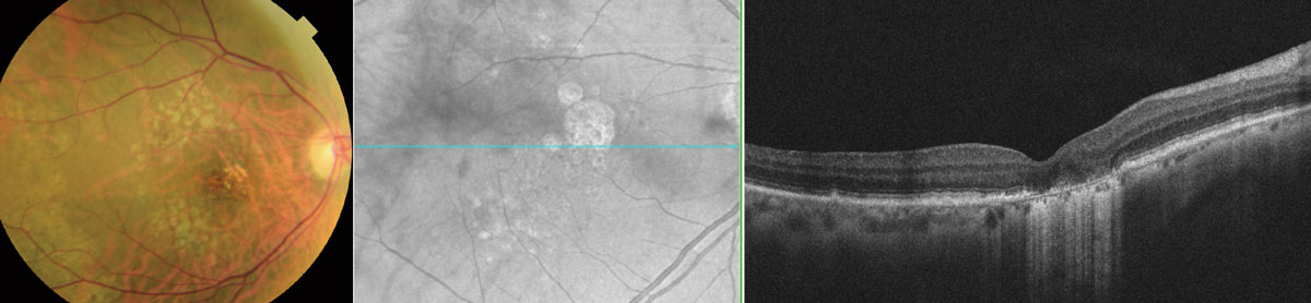
The presence of nascent GA serves as a significant risk factor for progression. Photo: Wendy Harrison, OD, PhD. Click image to enlarge. |
There is an urgent need to pinpoint disease biomarkers that could be used to identify high-risk individuals in the early stages of age-related macular degeneration (AMD). These biomarkers may also be used as earlier indicators of treatment efficacy and potentially serve as surrogate endpoints for geographic atrophy. After examining the association between the presence and development of incomplete retinal pigment epithelium (RPE) and outer retinal atrophy and the risk of subsequently developing geographic atrophy (GA), researchers recently noted that nascent GA was associated with a >80-fold increased risk of developing GA and explained >90% in the variance in time to develop GA proper.
Nascent GA is defined by the presence of a specific subset of the features of photoreceptor degeneration used to define incomplete RPE and outer retinal atrophy—the presence of the subsidence of both the outer plexiform layer and inner nuclear layer and/or the presence of the hyporeflective wedge-shaped band, without requiring evidence of choroidal signal hypertransmission or RPE attenuation or disruption needed in the latter.
The researchers performed OCT imaging and color fundus photography on 280 eyes from 140 participants with bilateral large drusen without defining features of nascent GA or late AMD at baseline and then at six-month intervals for up to 36 months. Eyes that developed neovascular AMD were censored the day it was detected. OCT volume scans were graded for the presence of incomplete RPE and outer retinal atrophy and nascent GA separately, and fundus photos were graded for the presence of GA.
The study confirmed that, when considered by itself, the presence and development of incomplete RPE and outer retinal atrophy was a significant risk factor for the subsequent development of GA. However, over a 24-month period, only 3% of these eyes developed GA. In contrast, 38% of eyes that developed nascent GA went on to develop GA over the 24-month study period. Furthermore, the proportion of variance in time to develop GA was significantly lower for incomplete RPE and outer retinal atrophy compared with nascent GA (43% compared with 91%, respectively). Incomplete RPE and outer retinal atrophy were no longer significantly associated with GA development when considered within the same model with nascent GA, suggesting that the risk of GA development may be mediated through its presence.
“The findings of this study provide evidence to support the notion that the anatomical features that define incomplete RPE and outer retinal atrophy can be considered a common risk factor in eyes with large drusen for the development of GA,” the researchers wrote in their paper in Ophthalmology. “Whilst replication is required through further studies, these findings potentially underscore the relatively greater utility of nascent GA as a surrogate endpoint compared with incomplete RPE and outer retinal atrophy.”
Wu Z, Goh KL, Hodgson LAB, Guymer RH. Incomplete retinal pigment epithelial and outer retinal atrophy: longitudinal evaluation in age-related macular degeneration. Ophthalmology. September 11, 2022. [Epub ahead of print]. |
