History

A 45-year-old black female presented for a routine eye examination. She had no ocular complaints, signs or symptoms. Her systemic history was remarkable for hypertensive medications. She had no pertinent ocular history and reported no allergies.
Diagnostic Data
Her best-corrected visual acuity measured 20/25 O.U. at distance and near. External examination was normal, with no evidence of an afferent pupillary defect. The anterior segment was normal and healthy.
Her intraocular pressure measured 19mm Hg O.U. The dilated fundus findings are illustrated in the photographs.
Your Diagnosis
How would you approach this case? Does this patient require any additional tests? What is your diagnosis? How would you manage this patient? What’s the likely prognosis?
Additional tests might include a Farnsworth Munsell 100 hue color vision test or panel D15 color vision test; Amsler grid testing; visual field testing to rule out functional deficits; and fundus photography. Sodium fluorescein angiography would not be indicated unless there was a reason to suspect choroidal neovascularization (CNV).
The diagnosis in this case is coloboma of both optic nerve heads. An ocular coloboma is defined as an area in the fundus that lacks tissue secondary to incomplete closure of the fetal embryonic fissure.1-8 Optic nerve colobomata (entities that are limited to the optic nerve) are described as part of the spectrum of large, abnormal-appearing optic discs where tissue is missing (usually inferonasally) that may be associated with other ocular colobomata.
In general, optic nerve coloboma (ONC) is a congenital condition that is typically discovered during initial fundus inspection or the first full eye examination. It may present unilaterally or bilaterally and can either mildly or severely affect visual function.1-4 ONC is often diagnosed in children who exhibit any or all of the following signs: afferent pupillary defect, loss of or decrease in color vision, strabismus, searching nystagmus and decreased visual acuity.1-6
The presence of decreased visual acuity varies greatly in documented cases of ONC and is dependant on the presence of damage to the fibers that subserve the fovea. Strabismus is not present in all cases of ONC. However, when present, strabismus is often caused by poor vision and sensory deprivation of the involved eye along with associated foveal hypoplasia or another issue pertaining to incomplete development. Furthermore, when strabismus is present, it often includes the phenomenon of searching nystagmus.3-8 Afferent pupillary defects produced by this anomaly may be highly pronounced, depending on laterality and severity of the disc excavation.1-6 The extent of color vision impairment is also dependant on the amount of missing tissue.1-6
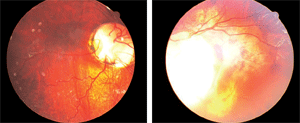
Dilated fundus images of our 45-year-old patient (O.D. left, O.S. right) reveal significant anatomical changes. What is your diagnosis?
Children who present with ONC and decreased visual acuity without frank strabismus should be evaluated for uncorrected refractive error. If a significant ametropia is absent and normal foveal anatomy is intact, occlusion therapy may be attempted to rehabilitate visual acuity.2
In many cases, ONC is accompanied by other ocular and/or systemic abnormalities, including malformations of the fundus and lens.3-8 Retinal detachments are common in ONC patients and create an uncertainty as to the overall ocular and functional prognosis.7,8
ONC eyes are also susceptible to serous macular detachments.3 Serous fluid is derived from one of three sources: fluid that perfuses into the retrobulbar space from surrounding orbital tissues; fluid from the peripapillary choriocapillaris; or cerebrospinal fluid.3
ONC is a common finding in reno-ocular syndrome.5 These patients have abnormalities of the thumb and renal system, lid ptosis, Duane’s retraction syndrome and ONC.5 ONC may also be associated with systemic dysfunction, including congenital heart defects and intracranial carotid artery anomalies.3-8
There is no treatment for ONC. Newly diagnosed patients must be examined for reno-ocular syndrome as well as other systemic and ocular malformations. Patients should be closely monitored for retinal and/or macular detachment with routine fundus examinations and home Amsler grid testing.
CHARGE syndrome is another entity composed of systemic issues in association with ocular coloboma.8 It consists of six major clinical features: coloboma of the eye, heart defects, atresia of the choanae, retarded growth and developmental anomalies (which include CNS anomalies), genital hypoplasia and/or urinary tract anomalies, and ear anomalies and/or hearing loss.8
This patient was photodocumented and educated regarding their unique findings. No additional treatment or work up was required.
Thanks to Virginia Donati, O.D., of Bolton, Ontario for contributing this case.
1. Lee BJ, Traboulsi EI. Update on the morning glory disc anomaly. Ophthalmic Genet. 2008 Jun;29(2):47-52.
2. Ari S, Keklíkçí U, Caça I, et al. Congenital isolate and total optic disc coloboma: case report and review of the literature. Ann Ophthalmol (Skokie). 2007 Spring;39(1):75-7.
3. Cekić S, Stanković-Babić G, Visnjić Z, et al. Optic disc abnormalities - diagnosis, evolution and influence on visual acuity. Bosn J Basic Med Sci. 2010 May;10(2):125-32.
4. Berk AT, Yaman A, Saatçi AO. Ocular and systemic findings associated with optic disc colobomas. J Pediatr Ophthalmol Strabismus. 2003 Sep-Oct;40(5):272-8.
5. Dureau P, Attie-Bitach T, Salomon R, et al. Renal coloboma syndrome. Ophthalmology. 2001 Oct;108(10):1912-6.
6. Parisi MA. Clinical and molecular features of Joubert syndrome and related disorders. Am J Med Genet C Semin Med Genet. 2009 Nov 15;151C(4):326-40.
7. Cheong HI, Cho HY, Kim JH, et al. A clinico-genetic study of renal coloboma syndrome in children. Pediatr Nephrol. 2007 Sep;22(9):1283-9.
8. Song JJ, Kwon SK, Cho CG, et al. Skull base vascular anomaly in CHARGE syndrome: a case report and review. Int J Pediatr Otorhinolaryngol. 2008 Apr;72(4):535-9
